42 white blood cells diagram
Before a transfusion, white blood cells are often removed to reduce the risk of infections or immune reactions. Looking at blood cells Many types of blood cell are 10 μm in size or less. 30 seconds. Q. White blood cells differ from red blood cells because only they contain _______. answer choices. a biconcave shape. a nucleus and most organelles. the ability to transport both oxygen and carbon dioxide. the iron-containing molecule called hemoglobin. Report Quiz.
Hi guys. Welcome back to my channel. In today's video, I will be sharing with you how to draw white blood cells at ease. Remember to subscribe to my YouTube ...

White blood cells diagram
2 Diagram of the human cell illustrating the different parts of the cell. Cell Membrane The cell membrane is the outer coating of the cell and contains the cytoplasm, substances within it and the organelle. It is a double-layered membrane composed of proteins and lipids. Structure: Tiny, irregular shaped cells with a diameter of 2-3 µm. Abundance: Present in less than 1% of the total blood cell count.Platelets are about 1/10th to 1/20th as abundant as white blood cells. Composition: Lacks nucleus and contains dense granules in the cytoplasm. Lifespan: 5 to 9 days Functions: Helping in blood clotting, the process of preventing hemorrhage in a damaged blood ... white blood cell diagram - Clinical Hematology Clinical Hematology Atlas, 3rd Edition Ideal for identifying cells at the microscope, this atlas covers the basics of hematologic morphology, including examination of the peripheral blood smear, basic maturation of the blood cell lines, and discussions of a variety of clinical disorders.
White blood cells diagram. Blood is a specialized body fluid. It has four main components: plasma, red blood cells, white blood cells, and platelets. Blood has many different functions, including: transporting oxygen and nutrients to the lungs and tissues. forming blood clots to prevent excess blood loss. carrying cells and antibodies that fight infection. 10 The diagram shows a cross-section through a plant stem. Q Q shows the part that is stained red when the stem is placed in water containing a red dye. What is found at Q? A guard cells B palisade cells C phloem D xylem 11 What could increase the rate of water uptake by a shoot? A covering the shoot with a black plastic bag White blood cells are the cells that help the body fight infection There are a number of different types and sub-types of white blood cells which each have different roles to play. The three major types of white blood cells are: Granulocytes Monocytes Lymphocytes Granulocytes There are three different forms of granulocytes: Neutrophils Eosinophils Basophils […] White Blood Cell Diagram. This human anatomy diagram with labels depicts and explains the details and or parts of the White Blood Cell Diagram. Human anatomy diagrams and charts show internal organs, body systems, cells, conditions, sickness and symptoms information and/or tips to ensure one lives in good health.
A white blood cell. White blood cells are the body's sentries, serving as the backbone of the immune system. White cells are found throughout the body, in both the blood and the lymphatic system. These blood cells have a density of about 4-11 billion per liter of blood. The scientific name for a white blood cell is leukocyte, simply meaning ... Leukopenia is a low white blood cell count that can be caused by damage to the bone marrow from things like medications, radiation, or chemotherapy. Folate or vitamin B12 deficiency can also result in it. So can lymphoma, in which cancer cells take over the bone marrow, preventing the release of the various types of white blood cells. HIV is ... Start studying 19-5 White Blood Cells. Learn vocabulary, terms, and more with flashcards, games, and other study tools. Download scientific diagram | Diagram of WBC (Eosinophil cell). from publication: White Blood Cell Nuclei Segmentation Using Level Set Methods and Geometric Active Contours | A new method for ...
Neutrophil Blood Cells; Lymphocyte Cell Diagram; White Blood Cell Components; Neutrophil Drawing; Neutrophil Cartoon; White Blood Cell Differential; White Blood Cells Histology; Phagocytic Cells; Dysplastic; White Blood Cell Types Chart; Under Microscope; 5 White Blood Cells; Lineage; High; Neutrophil Chemotaxis; Dendritic Cells and Macrophages ... Immune cell army is the immune system that protects human body against infection and pathogens. Illustration showing different types of cells in human circulatory system (white blood cells) and platelets. Modern illustration in high quality. blood cell diagram stock illustrations Think of white blood cells as your immunity cells. In a sense, they are continually at war. They flow through your bloodstream to battle viruses, bacteria, ... Find white blood cell diagram stock images in HD and millions of other royalty-free stock photos, illustrations and vectors in the Shutterstock collection.
The middle white layer is composed of white blood cells (WBCs) and platelets, and the bottom red layer is the red blood cells (RBCs). These bottom two layers of cells form about 40% of the blood. Plasma is mainly water, but it also contains many important substances such as proteins (albumin, clotting factors, antibodies, enzymes, and hormones ...
White blood cells (WBCs), or leukocytes, are immune system cells that defend the body against infectious disease and foreign materials. There are several different types of WBCs. They share commonalities but are distinct in form and function. WBCs are produced in the bone marrow by hemopoeitic stem cells, which differentiate into either ...
Elevated White Blood Cell Counts. Low White Blood Cell Counts. White blood cells (WBCs) are a part of the immune system. They help fight infection and defend the body against other foreign materials. Different types of white blood cells have different jobs. Some are involved in recognizing intruders.
Diagram White Blood Cells. Posted on June 5, 2014 by admin. Diagram White Blood Cells diagram and chart - Human body anatomy diagrams and charts with labels. This diagram depicts Diagram White Blood Cells. Human anatomy diagrams show internal organs, cells, systems, conditions, symptoms and sickness information and/or tips for healthy living.
Also called WBC and white blood cell. Blood cell development; drawing shows the steps a blood stem cell goes through to become ...
Start studying white blood cells. Learn vocabulary, terms, and more with flashcards, games, and other study tools.
18 The diagram shows the human heart. R P Q S In which order does blood pass through the chambers during a complete circuit of the body after it returns from the lungs? A Q → R → S → P B Q → R → P → S C P → S → Q → R D P → S → R → Q 19 An athlete takes part in a race.
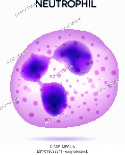
There are few organelles in the cytoplasm. Neutrophils. neutrophil diagram. Neutrophils are the commonest type of white blood cell found in a blood smear. They ...
Blood Tests. Complete blood count: An analysis of the concentration of red blood cells, white blood cells, and platelets in the blood. Automated cell counters perform this test.
White blood cells White blood cells of a human, photomicrograph panorama as seen under the microscope, 100x zoom. red blood cell diagram drawing stock pictures, royalty-free photos & images abnormal red blood cells abnormal red blood cells showing spherocyte.Medical science background concept. red blood cell diagram drawing stock pictures ...
Table 4.2 shows the number of white blood cells in the two blood samples. Table 4.2 white blood cells mean number of cells per mm3 of blood before infection during infection lymphocytes 1300 3500 phagocytes 2000 7500 ... The diagrams are not drawn to the same scale. Q P W S V R cell T microvilli
White Blood Cells (Leukocytes) White blood cells are much less numerous than RBCs (5,000 - 10,000 WBCs/µL blood) with a RBC/WBC ratio of approximately 700:1. WBCs work to protect the body from infection. WBCs are divided into two main groups based on cytoplasmic appearance: agranular leukocytes (lymphocytes and monocytes that have relatively ...
A white blood cell, also known as a leukocyte or white corpuscle, is a cellular component of the blood that lacks hemoglobin, has a nucleus, is capable of motility, and defends the body against infection and disease.White blood cells carry out their defense activities by ingesting foreign materials and cellular debris, by destroying infectious agents and cancer cells, or by producing antibodies.
White blood cells (WBCs), also called leukocytes or leucocytes, are the cells of the immune system that are involved in protecting the body against both infectious disease and foreign invaders. All white blood cells are produced and derived from multipotent cells in the bone marrow known as hematopoietic stem cells.Leukocytes are found throughout the body, including the blood and lymphatic system.
The white blood cells are also called Leukocytes. These cells act as a defence system against any infections in the human body. They produce special kinds of proteins called antibodies, which identify and fight pathogens invading the human body.These cells are classified further as granulocytes and agranulocytes.
White Blood cells. White blood cells or leukocytes (leukos = white, cytes = cells) are so-called because they are true cells that do not contain the red protein, hemoglobin.The real value of white blood cells is that most are specifically transported to areas of infection, thereby providing a rapid and potent defense against infectious agents. Normal count: the average total leukocytic count ...
Types of White Blood Cells. There are several types of wbcs, each one serving a unique goal. Let’s take a look at the leukocytes which take part in inflammatory disease conditions. They are essential parts of your immune system and can create a protection network for antigens coming from the environment.. Some of the white blood cells come from the bone marrow, while others come from the ...
White Blood Cell Amungst Red. Posted on September 10, 2014 by admin. White Blood Cell Amungst Red Diagram - White Blood Cell Amungst Red Chart - Human anatomy diagrams and charts explained. This diagram depicts White Blood Cell Amungst Red with parts and labels.
WBC-white blood cells are also called leukocytes or leucocytes. They are cells of the immune system, which is mainly responsible for: Protecting and fighting against invading pathogens. Stimulates the production of the progesterone hormone. Play a vital role in the human reproductive system by producing a network of blood vessels within the ovary.
white blood cell diagram - Clinical Hematology Clinical Hematology Atlas, 3rd Edition Ideal for identifying cells at the microscope, this atlas covers the basics of hematologic morphology, including examination of the peripheral blood smear, basic maturation of the blood cell lines, and discussions of a variety of clinical disorders.
Structure: Tiny, irregular shaped cells with a diameter of 2-3 µm. Abundance: Present in less than 1% of the total blood cell count.Platelets are about 1/10th to 1/20th as abundant as white blood cells. Composition: Lacks nucleus and contains dense granules in the cytoplasm. Lifespan: 5 to 9 days Functions: Helping in blood clotting, the process of preventing hemorrhage in a damaged blood ...
2 Diagram of the human cell illustrating the different parts of the cell. Cell Membrane The cell membrane is the outer coating of the cell and contains the cytoplasm, substances within it and the organelle. It is a double-layered membrane composed of proteins and lipids.


0 Response to "42 white blood cells diagram"
Post a Comment