41 identify which diagram represents cells that produce and circulate cerebrospinal fluid.
Cells . Cells are the smallest unit of life. To understand what a cell looks like, picture a chicken egg. It has an outer membrane (in the case of an egg, it's a hard shell, but most cells aren't like that); it's filled with nutrient-rich fluid (whites of the egg versus cytoplasm in a cell) and has a nucleus (egg yolk). Learn the ventricles of the brain along with their definition, function, location, anatomy, and cerebrospinal fluid (CSF) flow using labeled diagrams. The ventricular system contains the lateral, third, and fourth ventricles whose function is to produce cerebrospinal fluid. Learn where CSF is found,
Microvilli Definition. Microvilli, in the most simplistic terms, are tiny little microscopic projections that exist in, on, and around cells. They can exist on their own or in conjunction with villi (projections of some mucous membranes, most specifically of the small intestine, which are tiny folds that project out like numerous fingers).

Identify which diagram represents cells that produce and circulate cerebrospinal fluid.
Gram Staining Procedure/Protocol: Flood air-dried, heat-fixed smear of cells for 1 minute with crystal violet staining reagent. Please note that the quality of the smear (too heavy or too light cell concentration) will affect the Gram Stain results. Wash slide in a gentle and indirect stream of tap water for 2 seconds. Identify which diagram represents a cell that produces a myelin sheath in the ... diagram represents cells that produce and circulate cerebrospinal fluid. Rating: 5 · 2 reviews Convection - Process of heat exchange between the body and the surrounding air or fluid as a result of bulk flow of that air or fluid. Dehydration - Loss or deficiency of water in body tissues caused by sweating, vomiting or diarrhea. Symptoms include excessive thirst, nausea, and exhaustion.
Identify which diagram represents cells that produce and circulate cerebrospinal fluid.. Additionally, cerebrospinal fluid (CSF), produced in the four ventricular cavities of the brain, flows between the pia mater and arachnoid mater, providing protection from pathogens and mechanical support to the entire central nervous system. Special glial cells called ependymal cells produce CSF. The Brain They also produce cerebrospinal fluid and play a role in the Blood-brain barrier. Ependymal cells are incredibly tiny and form the surface by lining up firmly together. They possess cilia inside the ventricles, which resemble small hairs, moving in back and forth motion to circulate cerebrospinal fluid. Identify which diagram represents cells that produce and circulate cerebrospinal fluid. D. Identify the letter that indicates a Schwann cell. Ependymal cells, circulate cerebrospinal fluid and allow fluid exchange between the brain, spinal cord and CSF. Oligodendrocytes, insulate CNS axons Astrocytes, control chemical environment of neurons Microglial cells, protect CNS by scavenging dead cells and infectious microorganisms 24.
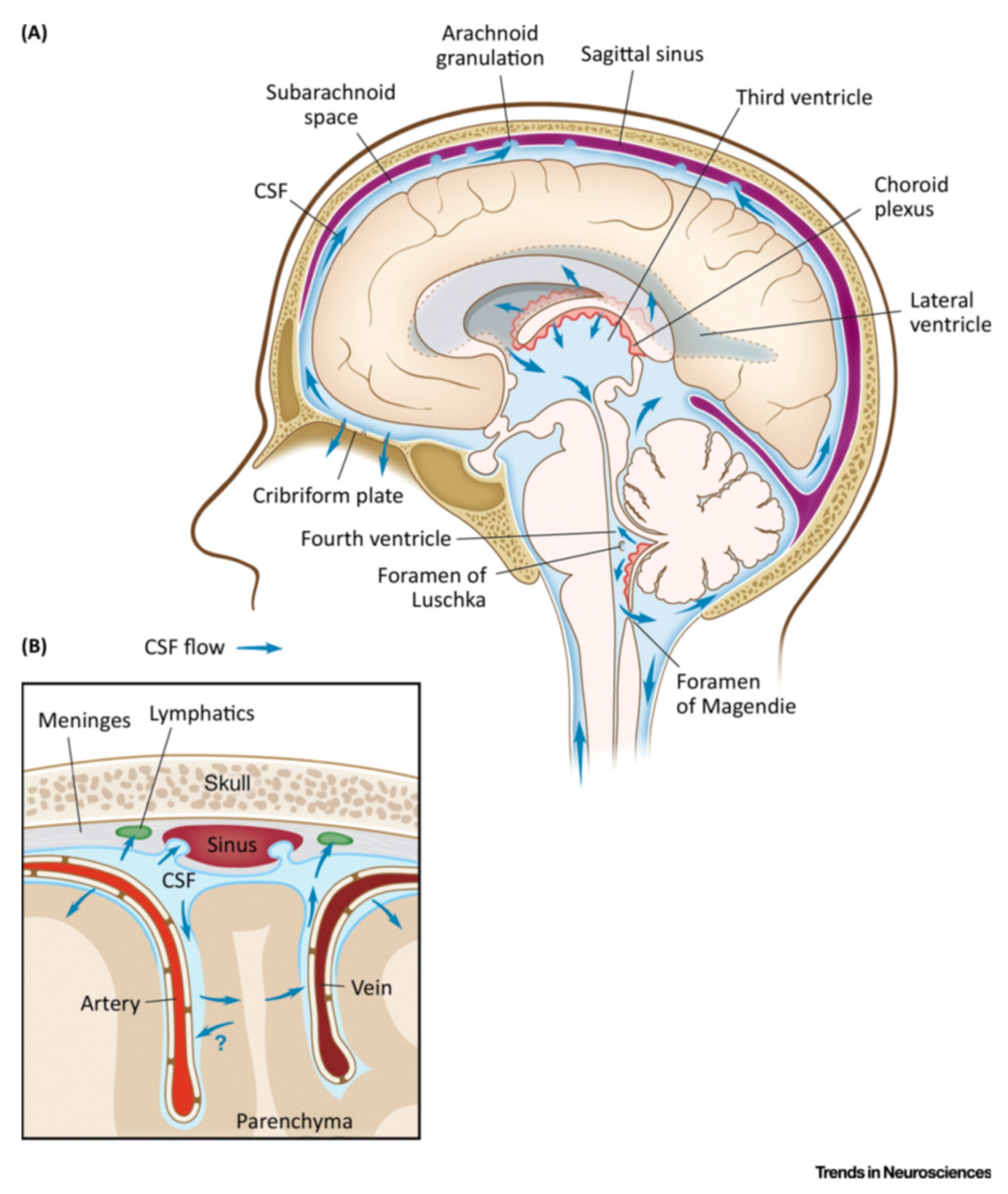
Stroke Volume. Stroke volume is defined as the difference between the volume of blood in the heart at the end of diastole (filling of the left ventricle) and the volume remaining in the heart at the end of systole- i.e. the volume of blood that is expelled with each heartbeat. Control of stroke volume is therefore directly related to the amount ... b) Help circulate cerebrospinal fluid. ... Identify which diagram represents cells that produce and circulate cerebrospinal fluid A B C D E. Ependymal cells, circulate cerebrospinal fluid and allow fluid exchange between the brain, spinal cord and CSF. Oligodendrocytes, insulate CNS axons Astrocytes, control chemical environment of neurons Microglial cells, protect CNS by scavenging dead cells and infectious microorganisms 24. Cerebrospinal fluid (CSF) is an ultrafiltrate of plasma contained within the ventricles of the brain and the subarachnoid spaces of the cranium and spine.[1] It performs vital functions, including providing nourishment, waste removal, and protection to the brain.[2] Adult CSF volume is estimated to be 150 ml, with a distribution of 125 ml within the subarachnoid spaces and 25 ml within the ...
Labeling, ranking, sorting, or sentence completion questions. All of these question types require you to position items into an area of the answer box. Answer these kinds of questions on a computer, not on a smartphone. Press Tab to move forward or Shift/Tab to move backwards through the provided answer items. Identify which diagram represents cells that produce and circulate cerebrospinal fluid. D. Image: Identify which diagram represents cells that produce and ... Neural pathways anatomy The central nervous system (CNS) contains numerous nerve fibers that group together to form pathways between its various parts. These neural pathways represent the communicating highways of the CNS. They can be located solely within the brain, providing connections between several of its structures, or they can link the brain and the spinal cord together. Identify which letter represents the most abundant category of glial cells in the CNS ... Ciliated neuroglial cells that form an epithelium and play an active role in forming and moving cerebrospinal fluid are • Ependymal cells. Any long axon is called a nerve fiber. ... Which letter indicates cells that produce melanin in the hair root? D.
Gastrulation is a process that creates three different germ layers in an early embryo called ectoderm, endoderm, and mesoderm. Learn what this process is and discover how it occurs in the ...
Identify which diagram represents a cell that produces a myelin sheath in the ... diagram represents cells that produce and circulate cerebrospinal fluid.
MBL pathway resembles classical pathway as it proceeds through the action of C4 and C2 to produce activated proteins of the complement system. MBL works same as C1q which it resembles in structure. After the MBL binds to carbohydrate residues on the surface of a cell or pathogen, two components, MASP-1 and MASP-2 bind to MBL.

Interstitial fluid is similar to blood plasma in many ways. Some of this fluid starts to flow into the extended open-ended network of tubular structures forming the lymphatic circulation. This fluid is now called lymph and passes through lymph nodes, where pathogens, damaged cells, or cancerous cells can be trapped and destroyed.
Nasal Cavity. The nasal cavity is a large, air-filled space in the skull above and behind the nose in the middle of the face. It is a continuation of the two nostrils. As inhaled air flows through the nasal cavity, it is warmed and humidified. Hairs in the nose help trap larger foreign particles in the air before they go deeper into the respiratory tract.

This group of cells is found between the atria and ventricles. It transmits electrical impulses from the atria, where the action potential is briefly delayed as they contract, to the AV bundle. The AV bundle (Bundle of His) The electrical connection between the atria and the lower chambers of the heart (the ventricles) is through the AV bundle.

Microglia are ciliated to help circulate cerebrospinal fluid (CSF). ... Identify which diagram represents cells that produce and circulate cerebrospinal ... Rating: 5 · 2 reviews
A: Schwann cells form myelin sheaths around axons, or enclose unmyelinated axons in the peripheral nervous system. B: Ependymal cells line ventricles of the brain, circulate cerebrospinal fluid; some form choroid plexuses, which produce CSF. D: Astrocytes serves as the major supporting tissue in the CNS and contribute to the blood-brain barrier. 6.

The human body contains five organs that are considered vital for survival. They are the heart, brain, kidneys, liver, and lungs. The locations of these five organs and several other internal organs are shown in Figure 10.4. 2. If any of the five vital organs stops functioning, the death of the organism is imminent without medical intervention.
Sympathetic nervous system (diagram) The autonomic system is made up of two divisions, the sympathetic and parasympathetic systemsThey usually work antagonistically in the organs, but in a well integrated manner. It is the balance of the actions of both divisions that maintains a stable internal environment in the body.

Microglia are ciliated to help circulate cerebrospinal fluid (CSF). ... Identify which diagram represents cells that produce and circulate cerebrospinal ...

Electroplating is widely used in industries such as automobile, airplanes, electronics, jewelry, and toys. The overall process of electroplating uses an electrolytic cell, which consists of putting a negative charge on the metal and dipping it into a solution that contains metal salt (electrolytes) which contain positively charged metal ions.
Organelles compartmentalize cells into discrete functioning units. Cells must regulate the number of organelles to achieve proper function (Marshall, 2007; Marshall, 2016; Nigg and Holland, 2018; Rafelski and Marshall, 2008).For example, multiciliated cells (MCCs) line the epithelia of the brain ventricles, the airway, and the oviduct where motile cilia propel extracellular fluid to circulate ...

Identify which diagram represents cells that produce and circulate cerebrospinal fluid. D.

Function. Aside from cerebrospinal fluid, your brain ventricles are hollow. Their sole function is to produce and secrete cerebrospinal fluid to protect and maintain your central nervous system. CSF is constantly bathing the brain and spinal column, clearing out toxins and waste products released by nerve cells.

The pressure in the cranial vault is measured in millimeters of mercury (mm Hg) and is normally less than 20 mm Hg. The cranium is a rigid structure that contains 3 main components: brain, cerebrospinal fluid, and blood. Any increase in the volume of its contents will increase the pressure within the cranial vault.
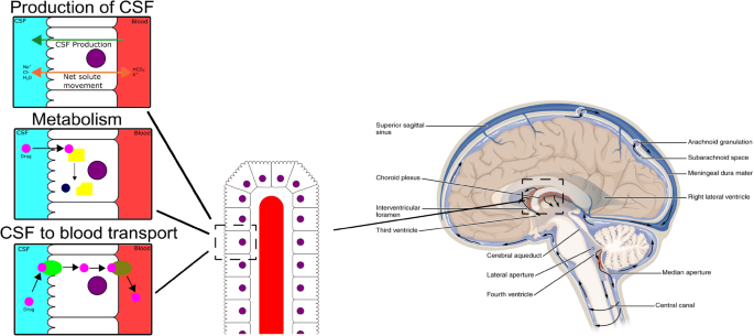
Cerebrospinal fluid circulation begins with the pulsing of the choroid plexus. Tiny cilia located on ependymal cells that also produce small amounts of CSF help propel the fluid along. It will eventually circulate throughout the subarachnoid spaces in the brain and spinal cord , and then be absorbed into the bloodstream.

Mantle Convection on Earth. Under the rigid layer of rock we live on, the Earth's asthenosphere is like dense plastic.. Because of its fluid-like properties, mantle convection can occur. Then, mantle convection is the main driver of plate tectonics.

The structure located in the ventricles that produces cerebrospinal fluid is called the choroid plexus. The cerebrospinal fluid is a clear, protective fluid made by the cells of the choroid plexus ...

Convection - Process of heat exchange between the body and the surrounding air or fluid as a result of bulk flow of that air or fluid. Dehydration - Loss or deficiency of water in body tissues caused by sweating, vomiting or diarrhea. Symptoms include excessive thirst, nausea, and exhaustion.

Identify which diagram represents a cell that produces a myelin sheath in the ... diagram represents cells that produce and circulate cerebrospinal fluid. Rating: 5 · 2 reviews
Gram Staining Procedure/Protocol: Flood air-dried, heat-fixed smear of cells for 1 minute with crystal violet staining reagent. Please note that the quality of the smear (too heavy or too light cell concentration) will affect the Gram Stain results. Wash slide in a gentle and indirect stream of tap water for 2 seconds.


0 Response to "41 identify which diagram represents cells that produce and circulate cerebrospinal fluid."
Post a Comment