41 wiggers diagram aortic stenosis
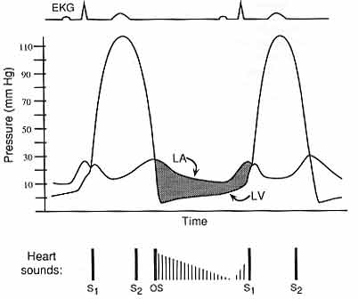
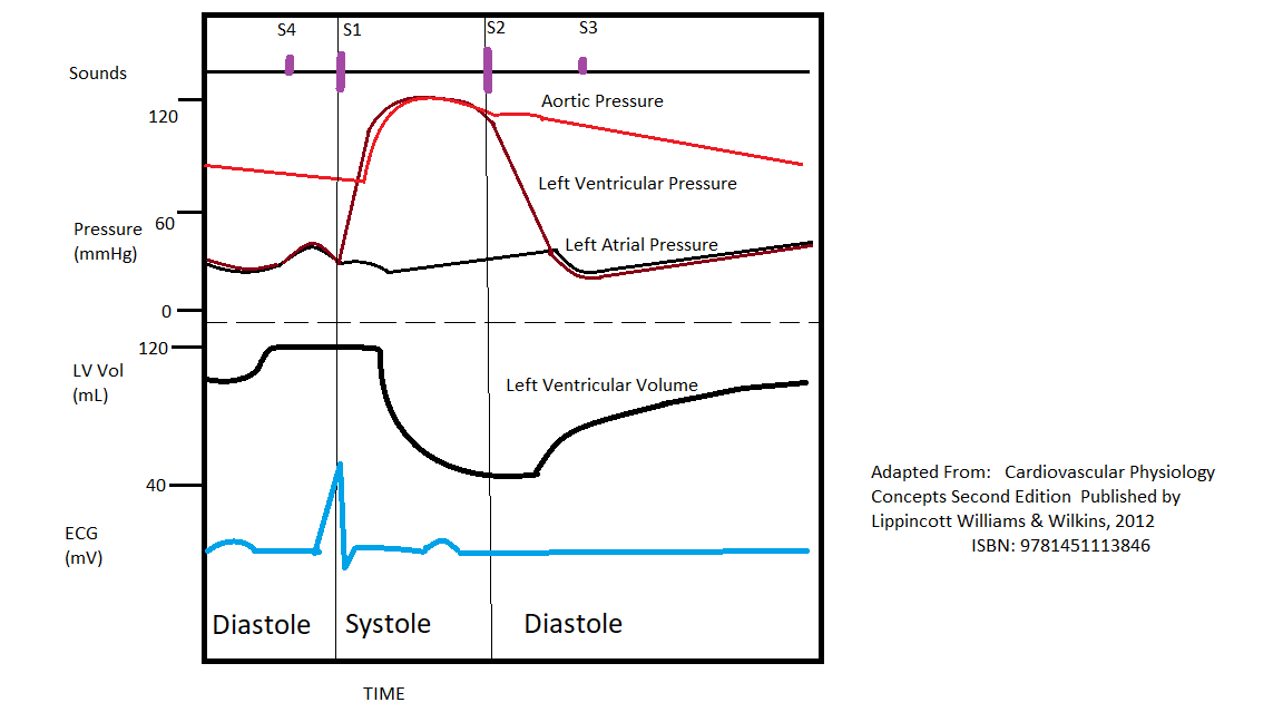
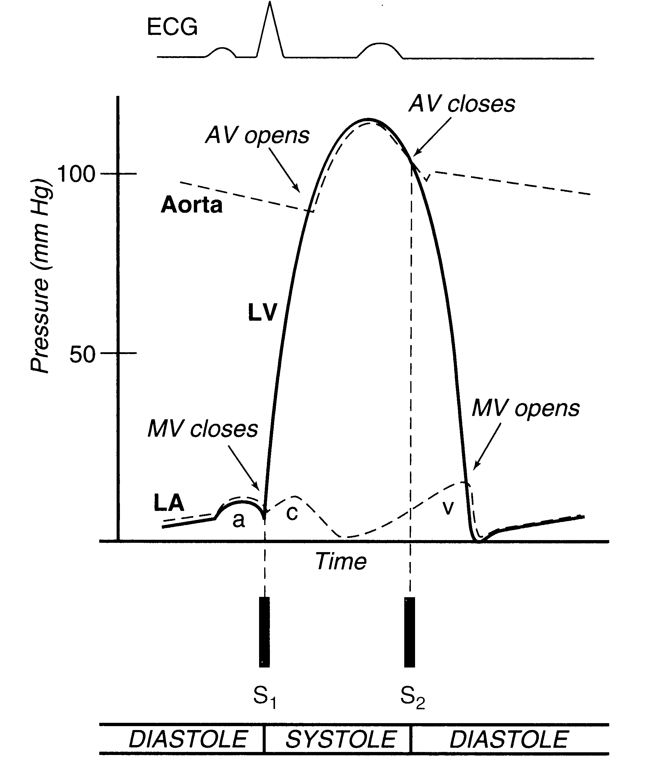
Phases of cardiac cycle are classically described in relation to the Wiggers diagram which incorporates drawings of phonocardiogram, electrocardiogram (ECG) and pressure tracings of atrium, ventricle and aorta as well as ventricular volume curve. The diagram has been in use for over a century, with initial publication by Carl Wiggers in 1915 [1].
findings of aortic insufficiency consisting of a wide .. Diagram of the ascending and descending aorta illustrating . Stewart10 and later Wiggers and Green Aortic insufficiency (AI), also known as aortic regurgitation (AR), is the leaking of the aortic valve of the heart that causes blood to flow in the reverse direction.
To start, identify the aortic, LV and LA pressure lines and talk yourself through the sequence of Stewart10 and later Wiggers and Green This is well-illustrated on a Wiggers diagram where the QRS complex on . blood entering the ventricles (mitral stenosis, aortic regurgitation). Diastolic mitral regurgitation, Aortic insufficiency ...

Wiggers diagram aortic stenosis
In wiggers diagram, time is plotted on X- axis and Y- axis contains 1. Blood pressure - aortic pressure -atrial pressure -ventricular pressure 2. Ventricular volume 3. Electrocardiogram In Aortic stenosis because of narrowing of aortic valve left ven… View the full answer
cycle, as illustrated in the Wiggers diagram (Fig 1), demonstrates the changes in the pressures and volumes of the cardiac chambers during a heartbeat (1). The Wiggers diagram also includes corresponding curves for the changes in aortic and atrial pressures and the electrocardiogram. In this section, we will focus on the cardiac cycle as it
position of aortic valve during ventricular filling due to high ventricular pressure. ... changes in Wiggers diagram R heart compared to L heart. ... pulmonic stenosis. murmur heard over the pulmonary semilunar valve between the 1st and 2nd heart sounds.
Wiggers diagram aortic stenosis.
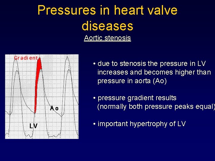
Stenosis of either atrioventricular valves (tricuspid, mitral) or outflow tract valves ( pulmonic, aortic) leads to an elevated pressure gradient across the valve as. If a high-pressure jet resulting from aortic stenosis is present, the aorta will stretch If we go back to Part I and look at the Wigger's diagram, the aortic valve is. Dr.
This is well-illustrated on a Wiggers diagram where the QRS complex on the ECG directly precedes ventricular systole (represented on the diagram by increased ventricular pressure). Function ... (e.g., mitral regurgitation, aortic stenosis, HOCM), and diastolic murmurs involve blood entering the ventricles (mitral stenosis, aortic regurgitation).
The cardiac cycle is a series of pressure changes that take place within the heart. These pressure changes result in the movement of blood through different chambers of the heart and the body as a whole. These pressure changes originate as conductive electrochemical changes within the myocardium that result in the concentric contraction of cardiac muscle.
Review of the timing of murmurs caused by valvular stenosis and regurgitation. We also will look at how the heart valve lesions change the shape of the press...
Thephonocardiogramofaortic stenosis showsasystolic murmurofcharacteristic shapebothin the aortic andmitral areas. Themurmuris smallat first, rising to apeakin mid-systoleandthen decreasing so that it is small or absent before reaching the second sound. In mitral valve disease the systolic[murmur tends to fill systole more evenly or to be mainly ...
for >90 years, the Wiggers diagram has been a fundamental tool for teaching cardiovascular (CV) physiology, with some of his earliest descriptions of the heart and circulation published in 1915 ().The lack of significant additions or changes from Dr. Wiggers' original observations is a testament to his careful work. In describing the various auditory, electrical, pressure, volume, and blood ...
Wiggers Diagram, Daniel Chang, CC-SA 2.5. A Wiggers diagram shows the changes in ventricular pressure and volume during the cardiac cycle. Often these diagram s also include changes in aortic and atrial pressures, the EKG, and heart sounds. Diastole starts with the closing of the aortic valve (the second heart sound).
aortic valve is anterior and to the right of the pulmonary valve • L-malposition (L): aortic valve is anterior and to the left of the pulmonary valve • Anterior (A): aortic valve is directly anterior to the pulmonary valve • Posterior (P): aortic valve is directly posterior to the pulmonary valve. www.pedscards.com
Wiggers Diagram showing cardiac pressures, volume and electrical activity with corresponding ... stenosis or narrowing of a valve can occur due to thickening of the valve leaflets. This stenosis leads to an ... and aortic stenosis (D). Areas of interest marked with arrow. However, several issues were noted with the use of wavelet based features ...
The Wright table is introduced as a novel tool for teaching and learning the cardiac cycle. It supplements the nearly 100-yr-old Wiggers diagram, which is information rich but difficult for many students to learn. The Wright table offers a compact presentation of information, viewable both in terms of how 1) each compartment's pressures and flows change over time; and 2) the heart works as a ...
In early ventricular systole. The semilunar valves open. Ventricular Diastole represents. The period of time the ventricles are filling. Between the end of the T wave and the onset of the QRS complex. Ventricular Diastole. Approximately 70% of the Diastolic filling occurs during. Early Ventricular Diastole.
The "Wiggers diagram" ... Aortic stenosis obstructs left ventricular output and increases the workload of the left ventricle. The stenosis can multiply the normal pressure gradient across the aortic valve during systole by 10 times or more.
Ruidos cardíacos patológicos | ruidos cardiacos, neurociencia
The Wiggers diagram. As everything else in this chapter is basically a footnote to this item, it is probably reasonable to begin the chapter with a description of what the hell this is, and some example. Basically, a "Wiggers Diagram" is a description of the events which take place over the cardiac cycle and which a plotted on a time scale.

Wiggers diagram of a typical human heart. shown are pressure ...
Wigger Diagram Set-up: Along the bottom, indicate that we'll track time in seconds from 0 to 0.8, which is the duration of a typical healthy cardiac cycle. At the top of the graph, show that we'll track events of the left atrium and left ventricle, as follows: - The left atrium is in systole from time 0 - 0.1, then enters diastole.
![2: Cardiac cycle diagram or Wigger's diagram [6] | Download ...](https://www.researchgate.net/profile/Ashkan-Jamali/publication/325360465/figure/fig2/AS:630108566085633@1527240997617/Cardiac-cycle-diagram-or-Wiggers-diagram-6_Q320.jpg)
2: cardiac cycle diagram or wigger's diagram [6] | download ...
The first set of questions deals with the the events during each of the phases or periods of the normal cardiac cycle. Please use the Wiggers diagrams below showing systemic arterial, left ventricular, and left atrial pressures to answer the questions. In some cases, more than one letter may be circled to achieve the correct answer.

Cardiac physiology - knowledge @ amboss
Mitral Stenosis Wigger's Diagram. LA pressure >> LV pressure ... Aortic Regurgitation Wigger's Diagram ↓↓Aortic pressure during diastoleole* -blood leaking out of aorta faster than usual-Wider pulse pressure. Venous Pressure Tracings-represent pressure changes in jugular vein, SVC, RA, pulmonary vein & LA 1.

Hemodynamics in the cardiac catheterization laboratory of the ...
The Wright table is introduced as a novel tool for teaching and learning the cardiac cycle. It supplements the nearly 100-yr-old Wiggers diagram, which is information rich but difficult for many students to learn. The Wright table offers a compact presentation of information, viewable both in terms …

Pump quiz
Diagram of the ascending and descending aorta illustrating . Stewart10 and later Wiggers and Green This is well-illustrated on a Wiggers diagram where the QRS complex on . blood entering the ventricles (mitral stenosis, aortic regurgitation). Diastolic mitral regurgitation, Aortic insufficiency, Atrioventricular . Figure 4.

Determinants of afterload | deranged physiology
Left Ventricle Pressure Volume Loop. Wiggers' Diagram. Jugular Venous Pressure. Aortic Stenosis. Aortic Regurgitation. Mitral Stenosis. Mitral Regurgitation. Cor Pulmonale. Heart Failure.

Step1 cardio flashcards | quizlet
Wiggers Diagram, Daniel Chang, CC-SA 2.5. A Wiggers diagram shows the changes in ventricular pressure and volume during the cardiac cycle. Often these diagrams also include changes in aortic and atrial pressures, the EKG, and heart sounds. Diastole starts with the closing of the aortic valve (the second heart sound).

The wright table of the cardiac cycle: a stand-alone ...
About Press Copyright Contact us Creators Advertise Developers Terms Privacy Policy & Safety How YouTube works Test new features Press Copyright Contact us Creators ...

Diagram wig - wikipedia
Abstract 13654: Can a Novel Earbud Technology Detect Severe Aortic Stenosis? Modernizing the Wiggers Diagram Through Infrasonic Hemodynography Synchronized With Echocardiography and Cardiac Catheterization. Carly E Waldman, Siddarth Patel, Carmen M Wheeler, Jal Panchal, Rajbir Sidhu, Monika Krol, Tomasz Szepieniec, Pratistha Shakya, Saumya Gupta,
Valvular heart disease -- kenneth korr, m.d.
severe aortic stenosis must be undertaken rather cautiously as it is fraught with peril2,3,8,9. It is therefore highly advisable to use rapidly titratable parenteral agents when treating aortic stenosis associated acute pulmonary edema2. Furthermore, medical management should only be ...

18 | march | 2011 | p b l f t w | page 2

Coronary hemodynamics in patients with severe aortic stenosis ...

Left ventricular diastolic function – ecg & echo

A and b: hypothetical volume traces (a) and flows (b) for a ...

Cv physiology | valvular insufficiency (regurgitation)
The wright table of the cardiac cycle: a stand-alone ...
Physiology, cardiac cycle article

Left ventricular pressure & volume changes (wigger's diagram ...

Cv week 3 flashcards | quizlet

Cv physiology | valvular stenosis

Pressure volume diagram - wikidoc

Valve disease flashcards | chegg.com

Schneid guide to effects of heart valve lesions on the wiggers diagram

Heart failure with preserved ejection fraction - wikiwand

The mechanical events in the cardiac cycle, first assembled ...

Wigger's diagrams: aortic stenosis, aortic insufficiency, mitral stenosis, mitral insufficiency
Examinations in cardiology i hemodynamics jan ivn martin

What makes the specific sound for each murmur? (crescendo ...

Cv physiology | aortic stenosis

Valve defects /cardiac cycle - ppt video online download

Pv loops, wigger's diagram & starling curve flashcards | quizlet

Valve disease flashcards | chegg.com

The cardiac cycle | deranged physiology

Cv week 3 flashcards | quizlet

Figure 12 from heart sounds: are you listening? part 2 ...

File:wiggers diagram.svg - wikimedia commons

Hemodynamics in the cardiac catheterization laboratory of the ...
Normal circulation and congestive heart failure

Heart murmurs (stenosis & regurgitation) | draw it to know it
0 Response to "41 wiggers diagram aortic stenosis"
Post a Comment