39 hip flexor anatomy diagram
Shoulder muscles : Anatomy and functions | Kenhub Feb 22, 2022 · Extrinsic muscles Trapezius The trapezius muscle is a large muscle that defines the nuchal region.It has three parts; descending, transverse and ascending. Same as in serratus anterior, the parts have different origins and insertions. Anatomy hip muscles Images, Stock Photos & Vectors ... Anatomy hip muscles images. 6,302 anatomy hip muscles stock photos, vectors, and illustrations are available royalty-free. See anatomy hip muscles stock video clips. Image type. Orientation.
Femur Anatomy, Diagram & Definition | Body Maps 20/01/2018 · The femur is the only bone located within the human thigh. It is both the longest and the strongest bone in the human body, extending from the hip to the knee.
Hip flexor anatomy diagram
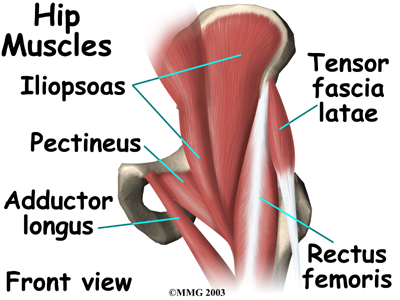
Hip Flexor Stretches to Counter the Effects of Sitting ... If done with proper alignment, Warrior Pose I can be a wonderful hip flexor stretch. Stand with one leg forward and one leg back, ready for Warrior I. Put your fingers on the front pelvis bones: You should be able to feel a small, round protuberance on each side, called the anterior superior iliac spine, or ASIS. Hip Muscles - Origin, Insertion, Action and Exercises ... Hip flexor muscles. These muscle flex the hip. Hip flexion is moving the leg forwards and upwards. The rectus femoris is also a hip muscle as well as being one of the quadriceps. Iliopsoas. Iliopsoas is sometimes classified as two muscles, Iliacus and Psoas major, with Iliacus arising from the Ilium and Psoas from the vertebrae. Leg Muscles: Anatomy, Support & Movement - Study.com 24/08/2021 · Leg muscles control movement of the foot and toes. Explore the anatomy, support, and movement of leg muscles including the extrinsic foot flexors and extensors, extrinsic toe flexors and extensors ...
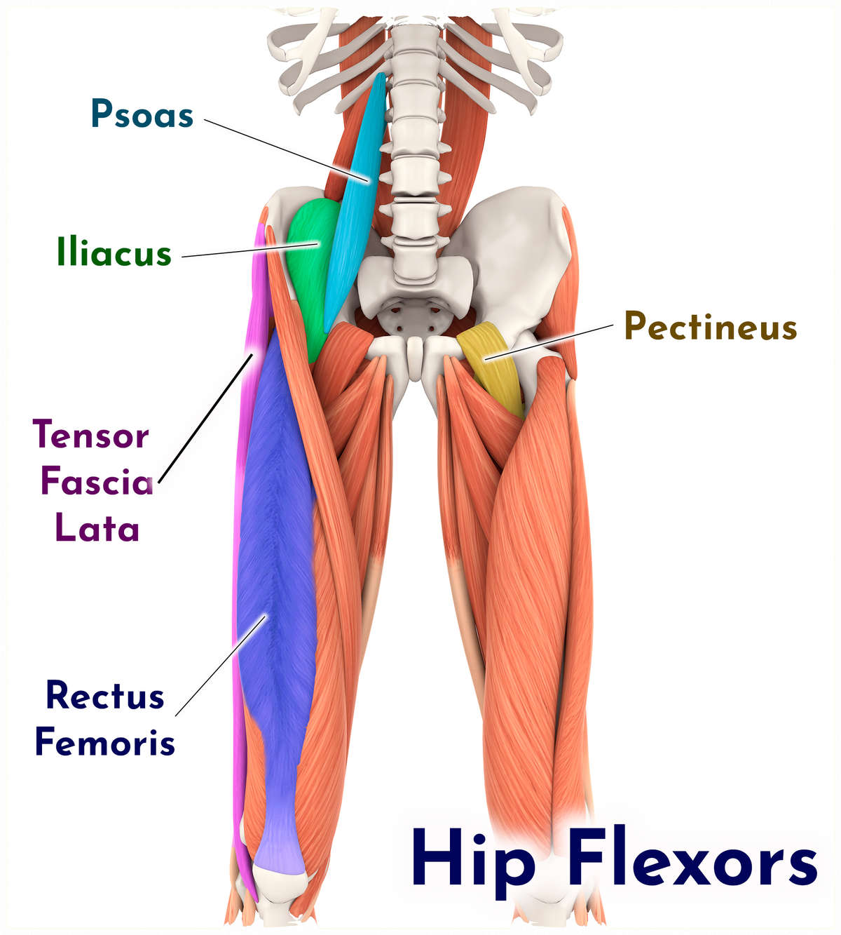
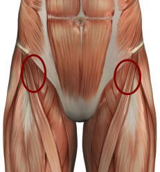
Hip flexor anatomy diagram. Anterior Hip Pain - Pain at the front of the hip 06/09/2019 · Explore the information under each tab below to understand more about the anatomy of the area and things that may go wrong. If you have already visited our Hip Pain Explained page, you may have already read some of this information. On this page you will find information specific to conditions related to pain in the front of the hip. Common conditions … Unlock Your Hip Flexors: HumanampAnimal Anatomy and ... Dec 2, 2017 - Unlock Your Hip Flexors: HumanampAnimal Anatomy and Physiology Diagrams le... Understanding Hip Flexor Pain - Sports-health Hip Flexor Muscles Anatomy. The hip flexor is not one singular muscle but a group of muscles, including the psoas major, iliacus, rectus femoris, pectineus, and sartorius. (The rectus femoris is also considered a quadricep muscle.) These muscles attach to various points on the spine, pelvis, and femur (thigh bone). Healing and Rehabbing Hip Flexor Tendonitis - NYDNRehab Hip flexor pain can be either acute, arising from trauma, or chronic, becoming worse over time. Common symptoms of hip flexor tendinosis include: Pain at the front of the hip or in the groin. Pain while walking or climbing stairs. Pain when lifting the knees toward the chest. Altered gait or limping due to hip or groin pain
Hip Muscles - The Definitive Guide | Biology Dictionary 27/06/2020 · Hip Flexor Exercises. You can exercise the hip flexor muscles sitting down. Sit straight and ensure your posture is good by imagining a string attached to the top of your skull that pulls your back upright. Both feet should face forward. Lift one knee so that the angle of the hip is above 90°. As the flexor muscles become stronger, you will be ... Hip Flexor Muscles and Anatomy for Personal Trainers All of the hip flexor muscles attach from the pelvis or spine to the femur or tibia, which is how they influence hip flexion. Bypass the tricky bony landmark terms for now and familiarize yourself with just the two bones each muscle attaches to. The bolded words in the descriptions below are there just for you intermediate anatomy student! ANATOMY - Medical Mnemonics .com ˜ From lateral hip towards medial navel: Nerve (directly behind sheath) Artery (within sheath) ... ˜ See inferior -view diagram. Knowledge Level 4, System: Cardiovascular Robert O'Connor University College Dublin Lung lobe numbers: right vs. left Hi Yield [ID 79] Tricuspid heart valve and tri-lobed lung both on the right side. Bicuspid and bi-lobed lung both on the left side. … What Are the 3 Main Hip Flexor Muscles? | Livestrong.com The hip flexor muscles are attached to the hip joint to allow the femur, which is the upper leg bone, to flex onto the pelvis region. In simpler terms, the hip flexor muscles allow the knee to raise and move the thigh upward. The hip is a large, deep and stable ball and socket joint that is surrounded by many ligaments, tendons and muscles.
CVM 6100 Veterinary Gross Anatomy Muscle names may be latinized (flexor digitorum profundus) or anglicized (deep digital flexor). Muscle are named (originally in the human) for their shape (deltoideus) or location (brachia-lis) or attachments (sternohyoideus) or structure (biceps) or function (supinator) or combinations of Diagram Of Hip Muscles And Ligaments | How To Unlock Your ... Iliopsoas tendinitis, in which hip flexor tendons end up being irritated, is one possibility presenting with inflammation and "snapping" in the hip socket. Pressure on the hip flexors can cause the muscles to tear, and this condition can range from minor to extreme depending on the level of the injury. Where Is Your Hip Flexor? - Body Pain Tips The Hip Flexor is not a single muscle in your body, but rather a combination of several muscles and tendons that are located in the hip region. All the muscles that are considered part of the Hip Flexor aid in hip flexion in one way or another. Hip flexion is the movement where your hip becomes contracted, most noticeably when you lift up your leg. Overview of Hip Flexor Muscles and Injuries The hip flexors are several muscles that bring your legs and trunk together in a flexion movement. They allow you to move your leg or knee up towards your torso, as well as to bend your torso forward at the hip. You can strain or tear your hip flexor muscles through sudden movements or falls. 1 Jan-Otto / E+ / Getty Images Anatomy and Function
Hip Flexor Muscles High Resolution Stock Photography and ...
A Guide to Hip Anatomy: Bones, Muscles, Tendons & Pain ... Adductor muscles on the inside of your thigh. Iliopsoas muscle, a hip flexor muscle that attaches to the upper thigh bone. Rectus femoris muscle, one of the quadriceps muscles on the front of your...

The Hip Flexor Complex | CMS Fitness Courses
CVM 6100 Veterinary Gross Anatomy Connective tissue structures identifiable in gross anatomy: Dermis [G. skin] — the physically tough/strong component of skin (deep to epidermis) Tendon — attaches muscle to bone (called aponeurosis when sheet-like) Ligament — attaches bone to bone (usually thickenings of fibrous joint capsules) [Note: visceral ligaments located in body cavities are entirely different …

Why Hip Flexors Are Tight and Why Your Hips Pop | Sparta Science
Understanding Hip Flexor Pains | What Causes Pain in Hip ... Avascular necrosis. This is a very rare hip flexor pain. It is caused when blood supply to a bone is completely cut off or significantly reduced. This type of hip pain is often felt in the center of the groin, thigh, or buttocks. It can develop gradually or can be caused by trauma, or excessive alcohol or steroid use.

How To Unlock Tight Hip Flexors - EMPOWER YOUR WELLNESS
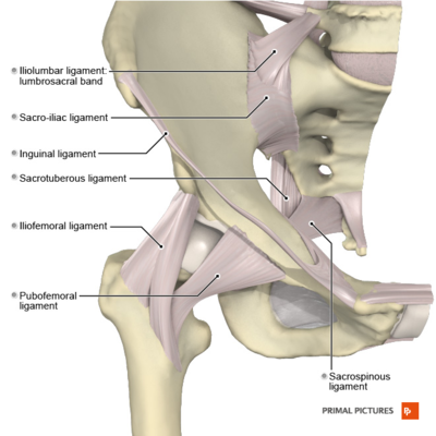
PDF Applied anatomy of the hip and buttock The flexor muscles of the hip joint (Table 1) are anterior to the axis of flexion and extension. pubofemoral ligament, in a more caudomedial orientation. Together they resemble the letter Z (Fig. 4). Posteriorly the capsule is strengthened by the ischiofemoral ligament (see Standring, Fig. 81.3).
Hip joint: Bones, movements, muscles | Kenhub
Skeletal System: Anatomy and Function, Diagram, Diseases, and ... Aug 30, 2018 · The human skeletal system consists of all of the bones, cartilage, tendons, and ligaments in the body. Altogether, the skeleton makes up about 20 percent of a person’s body weight.. An adult’s ...
Anatomy of the hip flexor musculature taken from Andersson ...
Shoulder muscles : Anatomy and functions | Kenhub 22/02/2022 · Muscles of the shoulder : Anterior view. The muscles of the shoulder support and produce the movements of the shoulder girdle.They attach the appendicular skeleton of the upper limb to the axial skeleton of the trunk. Four of them are found on the anterior aspect of the shoulder, whereas the rest are located on the shoulder’s posterior aspect and in the back.
توضيح Frail آلة حاسبة muscles of the leg and hip ...
Hip joint: Bones, movements, muscles - Kenhub The hip joint is a multiaxial joint and permits a wide range of motion; flexion, extension, abduction, adduction, external rotation, internal rotation and circumduction. Compared to the glenohumeral (shoulder) joint, however, this joint sacrifices mobility for stability as it is designed for weight bearing.
Hip Flexors Advanced Level – EasyFlexibility
Ankle and foot anatomy: Bones, joints, muscles | Kenhub Feb 22, 2022 · Ankle anatomy. The ankle joint, also known as the talocrural joint, allows dorsiflexion and plantar flexion of the foot.It is made up of three joints: upper ankle joint (tibiotarsal), talocalcaneonavicular, and subtalar joints.
Hip Strain | Symptoms & Treatment for Hip Flexor Strains | Buoy
Hip Muscles Diagram / Active Engagement (AE) Massage ... Back muscles diagram, body muscles diagram labeled, diagram of hip muscles and ligaments, hip anatomy diagram, hip muscles pain, thigh muscles diagram, human muscles. Source: massage.melbourne The diagram is a common one used to explain sliding filament theory, but don't worry about trying to the main muscles of the hip and pelvis consistsof ...
What is a Hip Flexor? - Plano Orthopedic & Sports Medicine Center
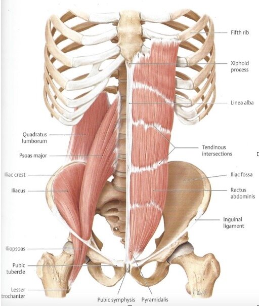
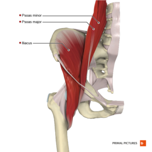
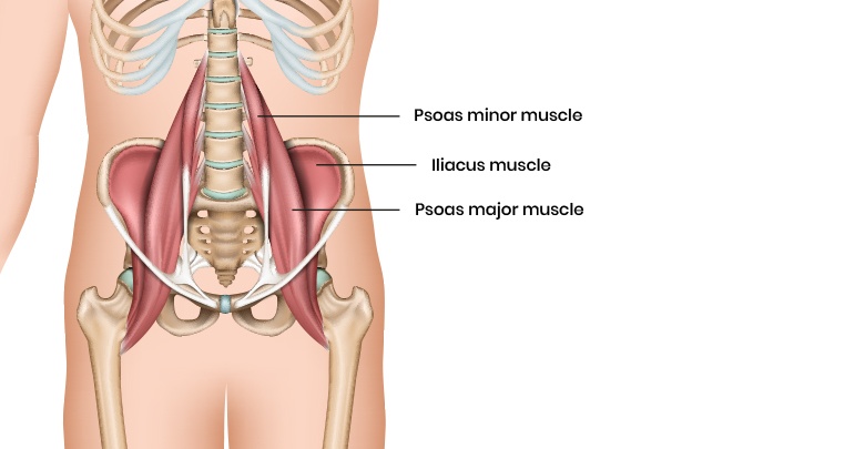
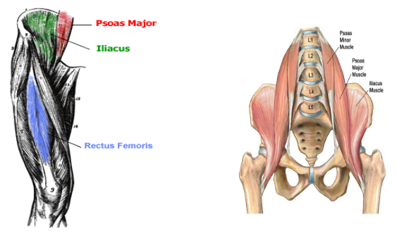
Anatomy of the Hip Flexor Muscles - Iliacus and the Psoas ... Anatomy of the Hip Flexor Muscles - Iliacus and the Psoas Major, Iliopsoas, Rectus Femoris Hip Flexor Muscle Anatomy The Iliopsoas actually consists of two muscles: the Iliacus and the Psoas Major. Together, they are known as the Iliopsoas. Anatomy Chart courtesy of FCIT The Iliacus originates on the pelvic crest and attaches on the femur.
Hip Flexors Stretch - How to Treat Your Hip Flexors | Girls ...
Basic Anatomy of Stretching the Hip Flexors - Movement Fix Live. •. This week we are returning to our discussion of 'the basic anatomy' series and we are taking a look at the hip flexors, but specifically the psoas and iliacus muscles. The hip flexors are all the muscles that flex the hip joint. Surprise! There are 4: sartorius, rectus femoris, iliacus, and psoas. Commonly the psoas and iliacus are ...

Psoas major muscle - Wikipedia
Hip Pain Explained - including structures & anatomy of the ... the joints of the hip & pelvis - hip joint, sacro-iliac joints, pubic symphysis the soft tissues - muscles, tendons, bursae & fascia bones - the femur (thigh bone) or bones of the pelvis local nerves running through and around the hip & pelvis musculoskeletal problems elsewhere in the body, such as the lower back (referred pain)
Hip Strains - OrthoInfo - AAOS
Femur Anatomy, Diagram & Definition | Body Maps Jan 20, 2018 · The femur is the only bone located within the human thigh. It is both the longest and the strongest bone in the human body, extending from the hip to the knee.
Hip Anatomy - Physiopedia
Patellofemoral pain syndrome (PFPS): a systematic review ... 26/06/2008 · Decreased hip flexor flexibility is assessed using the Thomas Test [74,83-85]. Weak hip abductors are evaluated using the Trendelenburg Test . A Q angle measurement in excess of 20 degrees may increase PFPS risk , however studies have demonstrated slight differences in Q angle between PFPS and control at lower Q angle values [55,57].
💪🏼💪🏿PT CORNER: Hip Flexor Pain 💪🏿💪🏼 — Fierce45®
Hip Diagram | How To Unlock Your Hip Flexors Hip Diagram Slide your left leg back until the top of your thigh rests on the ground. Utilizing your hands, gently push up until your spine is straight. To deepen the posture, place your lower arms on the ground and lean forward from your hips. Depending upon your versatility, you may be able to rest your forehead on the ground.

How To Unlock Tight Hip Flexors - EMPOWER YOUR WELLNESS
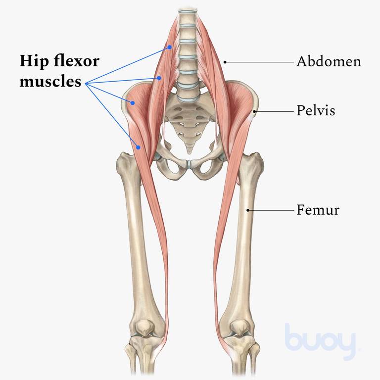
Hip flexor strain: Symptoms, recovery time, treatment, and ... The hip flexors connect the top of the femur, which is the largest bone in the body, to the lower back, hips, and groin. There are various hip flexor muscles that all work to enable a person to...

Tendinitis and Bursitis Treatment Cincinnati | Tendinitis ...
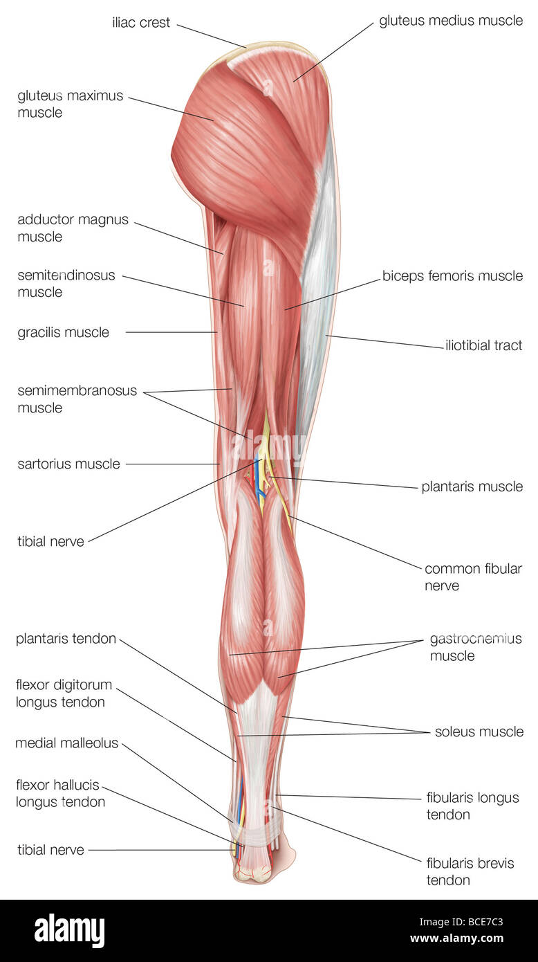
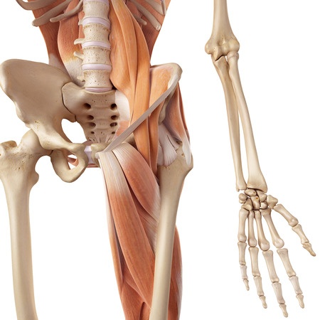
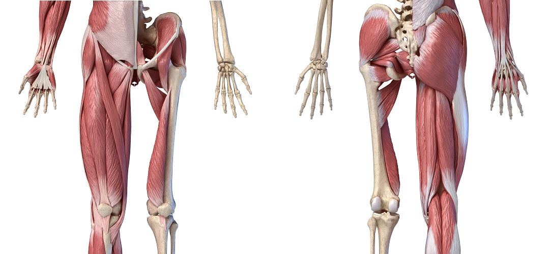
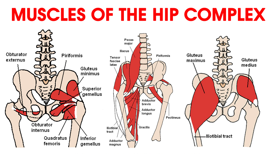
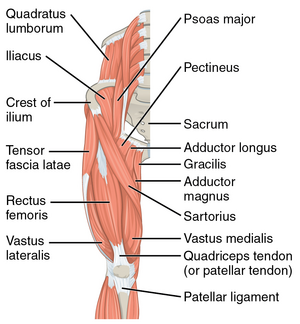
Muscles of the Hip - Anatomy Pictures and Information These muscles can be grouped based upon their location and function. The four groups are the anterior group, the posterior group, adductor group, and finally the abductor group. The anterior muscle group features muscles that flex (bend) the thigh at the hip. Continue Scrolling To Read More Below... Click To View Large Image Continued From Above...
It's All in the Hips: Part 3 - Athletico
Hip and thigh muscles: Anatomy and functions | Kenhub Small and deep muscles which mainly externally rotate the thigh at the hip joint and stabilize the pelvis. These are the piriformis, obturator internus, obturator externus, gemellus superior, gemellus inferior, and quadratus femoris. They are also known as the inner hip muscles and deep external rotators. Superficial layer

Hip Flexor - Home | Facebook
Cross sectional anatomy - Kenhub 20/03/2022 · Cross section through the maxillary sinus: Diagram There is no hidden agenda with regards to orientation, so it’s as easy as it gets. The brain (namely the brainstem and the cerebellum) points posteriorly (bottom of the image) and as you know from anatomy, the skull bones containing the paranasal sinuses are located anteriorly (top of the image).
Physical Therapy in Lake Forest for Hip - Anatomy
Iliopsoas Muscle: Anatomy, Function, and Treatment The iliopsoas muscle is a major mover of your hip joint. It's formed by the joining of three muscles: the iliacus muscle, the psoas major muscle, and the psoas minor muscle. These muscles work together to flex your hip and to stabilize your hip and lower back during activities such as walking, running, and rising from a chair.

Hip Flexor Strain – Considerations and Understanding
Hip Biomechanics - Recon - Orthobullets (OBQ09.172) Figure A represents a free body diagram of the hip of a patient standing on the right leg. The forces and distances are labeled on the diagram and the resulting hip joint force (J) = 1800N.

Deep Dive into the Anatomy of the Hip Flexor Muscles
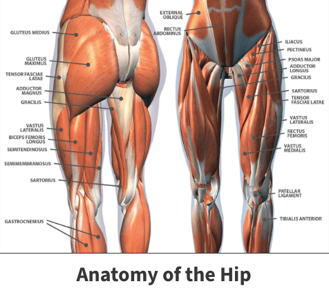
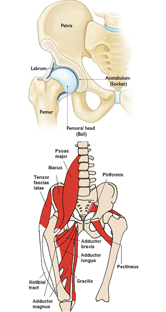
Hip Anatomy Diagram: From Bones To Joints - Science Trends The bones found in the hip include the three bones of the pelvis and the thigh bone or femur. The three bones which form the pelvis are the pubis, the ilium, and the ischium. The ilium is the bone at the top of the waist, while the pubis bones are found just below the ilium. The pubis curves downward and forwards from the ileum.
Hip Flexor Muscles and Anatomy for Personal Trainers
Hip Anatomy - Physiopedia The hip joint connects the lower extremities with the axial skeleton. The hip joint allows for movement in three major axes, all of which are perpendicular to one another. The location of the center of the entire axis is at the femoral head. The transverse axis permits flexion and extension movement.

Hip Pain Explained - including structures & anatomy of the ...
human muscle system | Functions, Diagram, & Facts | Britannica human muscle system, the muscles of the human body that work the skeletal system, that are under voluntary control, and that are concerned with movement, posture, and balance. Broadly considered, human muscle—like the muscles of all vertebrates—is often divided into striated muscle (or skeletal muscle), smooth muscle, and cardiac muscle. Smooth muscle is under involuntary control and is ...

Hip Flexors & Lateral Tight Diagram | Quizlet
18 Tips for Bulletproof Knees | Muscle diagram, Quad ... Hip Flexor Pain. Tight Hip Flexors. Biceps Femoral. Muscle Diagram. Muscle Anatomy. More information... . More like this. Yoga Anatomy. Anatomy Study. Soleus Muscle. Gastrocnemius Muscle. Medical Mnemonics. Achilles. The Secret. Health ... The Shoulder and Elbow Anatomical Chart illustrates general shoulder and elbow anatomy. The Shoulder and ...
Hip Flexors - Physiopedia
Hip Anatomy, Pictures, Function, Problems & Treatment The hip joint is a ball-and-socket type joint and is formed where the thigh bone (femur) meets the pelvis. The femur has a ball-shaped head on its end that fits into a socket formed in the pelvis, called the acetabulum. Large ligaments, tendons, and muscles around the hip joint hold the bones (ball and socket) in place and keep it from dislocating.

Hip Flexor Pain
Leg Bones Anatomy, Function & Diagram | Body Maps 30/03/2015 · The femur, or thighbone, is the longest and largest bone in the human body. At its top, it helps create the ball-and-socket joint of the hip; its …
Hip Muscles - The Definitive Guide | Biology Dictionary
Leg Muscles: Anatomy, Support & Movement - Study.com 24/08/2021 · Leg muscles control movement of the foot and toes. Explore the anatomy, support, and movement of leg muscles including the extrinsic foot flexors and extensors, extrinsic toe flexors and extensors ...

Hip Flexor Treatment in Newcastle | Your Trusted Physio
Hip Muscles - Origin, Insertion, Action and Exercises ... Hip flexor muscles. These muscle flex the hip. Hip flexion is moving the leg forwards and upwards. The rectus femoris is also a hip muscle as well as being one of the quadriceps. Iliopsoas. Iliopsoas is sometimes classified as two muscles, Iliacus and Psoas major, with Iliacus arising from the Ilium and Psoas from the vertebrae.

Six rotation movements on the X, Y, and Z axes of the hip ...
Hip Flexor Stretches to Counter the Effects of Sitting ... If done with proper alignment, Warrior Pose I can be a wonderful hip flexor stretch. Stand with one leg forward and one leg back, ready for Warrior I. Put your fingers on the front pelvis bones: You should be able to feel a small, round protuberance on each side, called the anterior superior iliac spine, or ASIS.

Iliopsoas - Wikipedia
Hip Flexor Exercise - Stretch for tight hip flexor muscles Oh ...

Probably more vulnerable - The Norway News

Deep Dive into the Anatomy of the Hip Flexor Muscles
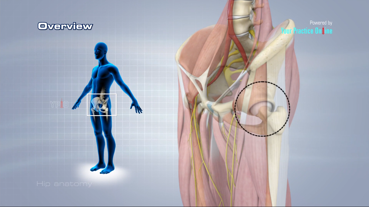
Hip Anatomy Video | Hip Orthopaedics Videos | Your Practice ...
Ask the Physio … Hip Flexors - Dance Life

Tendinitis and Bursitis Treatment Cincinnati | Tendinitis ...
Hip Flexors - Physiopedia
5 Hip Flexor Stretches!
0 Response to "39 hip flexor anatomy diagram"
Post a Comment