39 spinal cord cross section diagram labeled
Spinal Cord Cross Section Labeling - Printable This is a free printable worksheet in PDF format and holds a printable version of the quiz Spinal Cord Cross Section Labeling. By printing out this quiz and taking it with pen and paper creates for a good variation to only playing it online. This printable worksheet of Spinal Cord Cross Section Labeling is tagged. Click on the tags below to ... Spinal Cord Diagram Unlabeled - Wiring Diagram Pictures The spinal cord grey matter and the fiber tracts of the white matter.Answers from trusted physicians on labeled diagram of spinal cord. First: The spinal cord is roughly 18 inches long and somewhere between 1/4 and 1/2 inch in diameter.
Learn the spinal cord with diagrams and quizzes | Kenhub Take a look at the spinal cord cross section diagram below. Here you can see the white and gray matter of the spinal cord and the associated structures such as funiculi, lamina and tracts. Thinking of the information you learned in the video, spend some time linking the location of the labeled structures with what you know about their function.
Spinal cord cross section diagram labeled
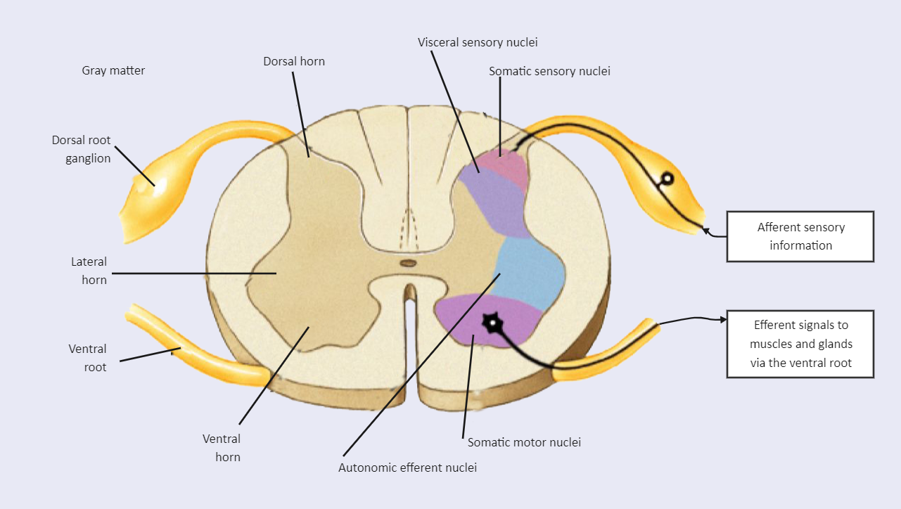
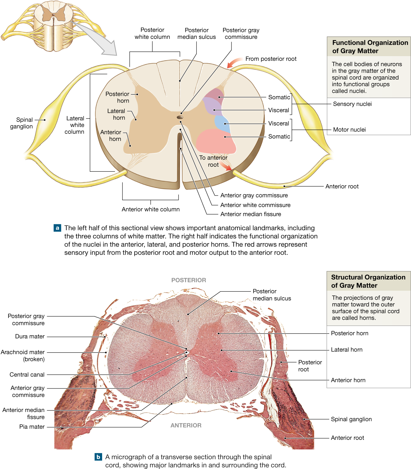
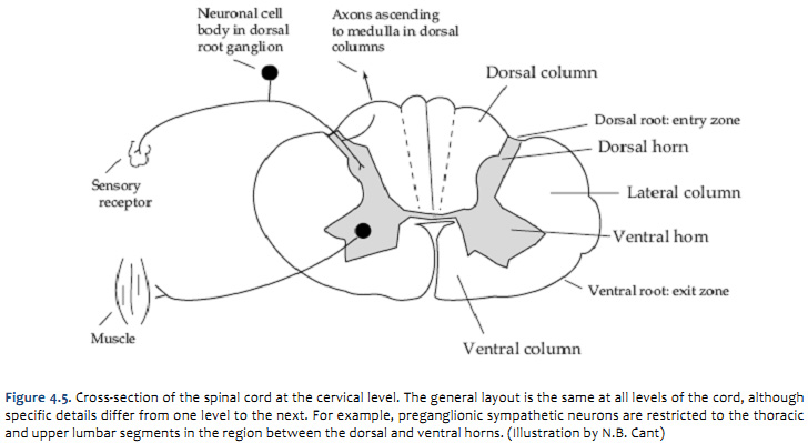
Spinal Cord Cross Section Labeled | EdrawMax The following diagram shows the spinal cord of the human body. In the spinal cord cross-section labeled diagram, we have shown Dorsal horn, Visceral sensory nuclei, Somatic sensory nuclei, afferent sensory information, efferent signals to muscles and glands via the ventral root, somatic motor nuclei, autonomic efferent nuclei, ventral horn ... Spinal Cord Quiz: Cross-Sectional Anatomy - GetBodySmart Spinal Cord - Cross-Sectional Anatomy. Start Quiz. Want to learn faster? Look no further than these interactive, exam-style anatomy quizzes. Learn anatomy faster and remember everything you learn. Start Now. Related Articles. Parts of the Brain Quiz. Test your knowledge with the parts of the brain and their functions in a fun and interactive ... Duke Neurosciences - Lab 2: Spinal Cord & Brainstem ... A cross-section through the spinal cord is illustrated schematically in Figure 2.6 and 3.4. The gray matter forms the interior of the spinal cord; it is surrounded on all sides by the white matter. The white matter is subdivided into dorsal (or posterior), lateral, and ventral (or anterior) columns.
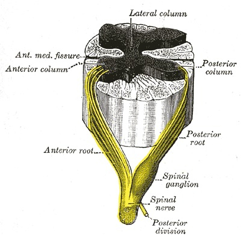
Spinal cord cross section diagram labeled. Solved 1. For the diagram of a spinal cord cross section ... Label: White matter, posterior and anterior gray horns, anterior median fissure, posterior median sulcus c. Draw and label: Question: 1. For the diagram of a spinal cord cross section below, sketch and label the following structures and nervous system subdivision: (3 points) a. PDF Anatomy of The Spinal Cord- Cross Section different parts of the spinal cord and between the cord and the brain. Focus on a small area of white matter using high magnification. View and draw a small area of the white matter. Include SEVERAL cross-sections of mostly axons of the neurons. Label the axon and myelin sheath on one of the cross sections. 4. Spinal Cord | ClipArt ETC Spinal Cord. "Diagram of a cross-section of the spinal cord through the roots of spinal nerves. c, central canal; d.f., dorsal fissure; d.r., dorsal root of spinal nerve arising from the dorsal horn of the gray matter (g); gn., ganglion on the dorsal root; n, spinal nerve; v.f., ventral fissure; v.r., ventral root of the spinal nerve, arising ... Cross-sections of the Spinal Cord - SmartDraw Cross-sections of the Spinal Cord Create healthcare diagrams like this example called Cross-sections of the Spinal Cord in minutes with SmartDraw. SmartDraw includes 1000s of professional healthcare and anatomy chart templates that you can modify and make your own. 24/75 EXAMPLES EDIT THIS EXAMPLE Text in this Example:
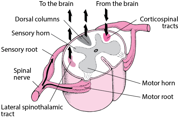
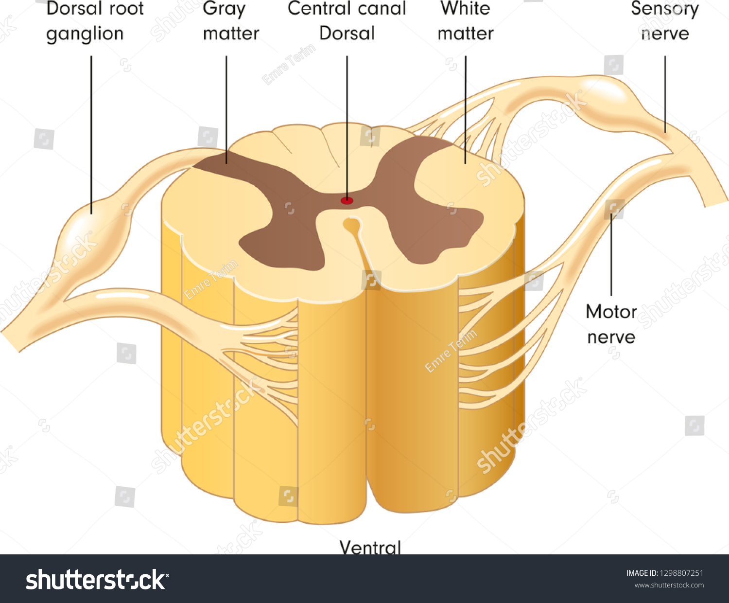
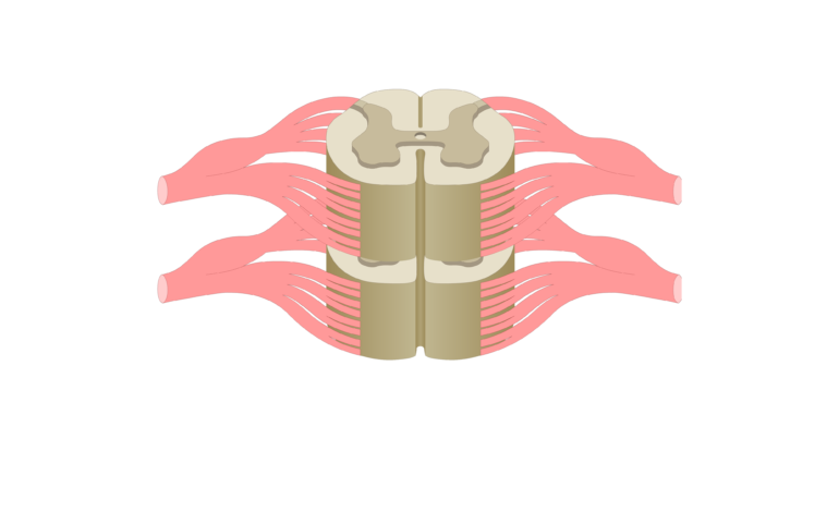
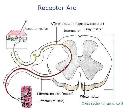
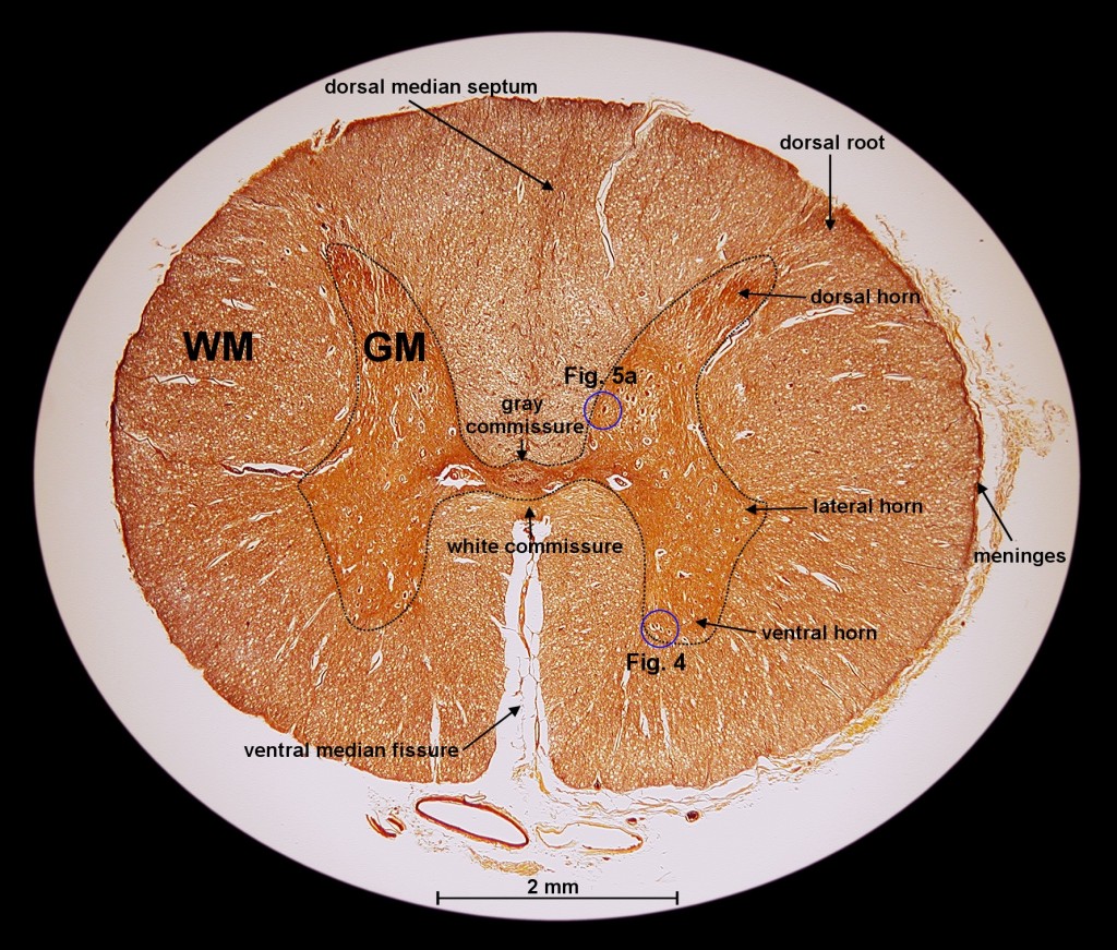
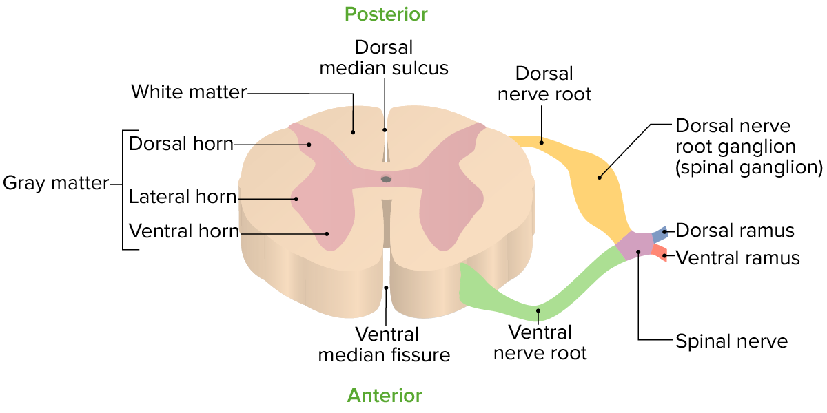
Spinal cord: Anatomy, structure, tracts and function | Kenhub The spinal cord is made of gray and white matter just like other parts of the CNS. It shows four surfaces: anterior, posterior, and two lateral. They feature fissures (anterior) and sulci (anterolateral, posterolateral, and posterior). The gray matter is the butterfly-shaped central part of the spinal cord and is comprised of neuronal cell bodies. Solved Spinal Cord and Reflex Arc Label the following ... Spinal Cord and Reflex Arc Label the following parts of a spinal cord on the cross-section diagram a. white matter b. grey matter c. dorsal root ganglion d. nerve fibers e. interneuron f. synapse g. sensory neuron h. motor neuron Reflex Act the following parts of a reflex are on the diagram of a person stepping on a tack and jerking their leg ... Cross-Sectional Anatomy - The Central Nervous system Cross-sectional anatomy of the spinal cord. The spinal cord appears to be somewhat flat with two grooves that mark its surface. The two grooves are named as follows: the ventral (anterior) median fissure and the more shallow dorsal (posterior) median sulcus. These two grooves run the length of the cord and partially divide it into right and ... Spinal Cord Cross Section Explained (with Videos) | New ... Spinal Cord Cross Section Looking at a cross section of the spinal cord, you would see gray matter shaped like a butterfly surrounded by white matter. The gray matter is the core and ends up to be four projections that are known as horns. At the back are two dorsal horns and away from the back are two ventral horns.
PDF Scanned Document - Bronx High School of Science SPINAL CORD AND REFLEX ACT Cross Section of Spinal Cord Label the following parts of a spinal cord on the cross-section diagram. Name a, b. c. d. h, white matter grey matter dorsal root ganglion nerve fibers interneuron synapse sensory neuron motor neuron (à)çpjnal cord newon. neuwn Spinal cord - austincc.edu Spinal Cord 40X Cross sections of the spinal cord are so large that you will not be able to see the whole thing on the microscope--you will have to move back and forth or use a dissecting microscope. This section is from a slide that includes both the spinal cord and a vertebra. Leave a Comment Cancel reply - BYJUS Spinal Cord Diagram Spinal Cord Diagram The spinal cord is one of the most important structures in the human body. In fact, it is the most important structure for any vertebrates. Anatomically, the spinal cord is made up is made up of nervous tissue and is integrated into the spinal column of the backbone. Main Article: Spinal cord cross section (Gray's illustration ... You can use Radiopaedia cases in a variety of ways to help you learn and teach. Add cases to playlists; Share cases with the diagnosis hidden
Solved] Label the following spinal cord cross section diagram ...
Spinal cord - Wikipedia The spinal cord is a long, thin, tubular structure made up of nervous tissue, which extends from the medulla oblongata in the brainstem to the lumbar region of the vertebral column. It encloses the central canal of the spinal cord, which contains cerebrospinal fluid. The brain and spinal cord together make up the central nervous system (CNS).
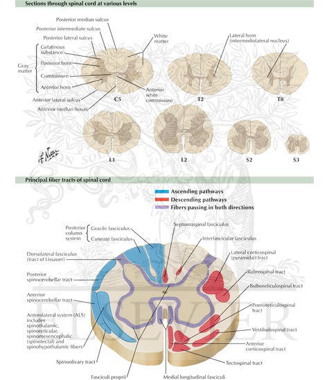
Spinal Cord Cross Sections: Fiber Tracts
The spinal cord | Human Anatomy and Physiology Lab (BSB 141) The spinal cord in cross-section has a central region of darker gray matter and the rest is lighter white matter. The gray matter is made up of neuroglia cells and neuron cell bodies. The white matter is made up of neuron axons, mostly but not all myelinated. The dorsal horns are the thinner projections of dark matter that jut out from the rest ...

Alila Medical Media | Cross section of the spinal cord ...
Spinal Cord Anatomy - Parts and Spinal Cord Functions A spinal needle is inserted between two vertebrae at level L3/L4 or L4/L5, where there is no risk of accidental injury to the spinal cord (which ends at L1 to L2). Cross-Sectional Anatomy of Spinal Cord. The spinal cord, like the brain, consists of two kinds of nervous tissue called gray and white matter.

Cross Section through Spinal Cord Diagram | Quizlet
Spinal Cord Cross Section Stock Photos, Pictures & Royalty ... View spinal cord cross section videos Browse 2,483 spinal cord cross section stock photos and images available, or search for spinal cord nerve or spinal cord injury to find more great stock photos and pictures. Newest results spinal cord nerve spinal cord injury neurons central nervous system cardiac muscle biomedical illustration
90053508 Style Medical 1 Nervous 1 Piece Powerpoint ...
Spinal cord cross section (Gray's illustration ... Some of Gray's illustrations of the spinal cord.

The spinal cord | Human Anatomy and Physiology Lab (BSB 141)
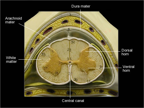
Spinal cord- Cross section labeled w/ functions Diagram ... on each side of the spinal cord between the anterior and posterior columns Posterior (dorsal) column lie between the posterior horns and posterior median sulcus Pia mater "delicate mother" inner layer of cord Arachnoid mater spidery looking middle layer; carries blood vessels etc. Subarachnoid space filled with CSF (spinal taps) Dura mater

Nerve Root Anatomical Structure Labeled Cross Section Stock ...
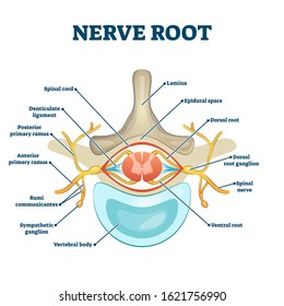
PDF In the diagram above label the following in the diagram above clearly label the following: 1) dorsal (or posterior) root 2) dorsal root ganglion 3) ventral (or anterior) root 4) dorsal horn 5) ventral horn 6) axon of incoming senory neuron 7) cell body of motor neuron 8) spinal cord interneuron 9) central canal of spinal cord 10) spinal cord white matter 11) spinal cord gray matter 12) …

The sketch of the cross section of the spinal cord connected ...
Spinal Cord Cross Section Illustrations, Royalty-Free ... Browse 1,080 spinal cord cross section stock illustrations and vector graphics available royalty-free, or search for spinal cord nerve or spinal cord injury to find more great stock images and vector art. Newest results spinal cord nerve spinal cord injury neurons central nervous system cardiac muscle biomedical illustration

Solved: Chapter 13 Problem 20SAE Solution | Human Anatomy 8th ...
Spinal Cord - Anatomy, Structure, Function, & Diagram Cross-section of spinal cord displays grey matter shaped like a butterfly surrounded by a white matter. Grey matter consists of the central canal at the centre and is filled with a fluid called CSF (Cerebrospinal fluid). It consists of horns (four projections) and forms the core mainly containing neurons and cells of the CNS.
Spinal Cord Cross Section Labeled | EdrawMax
Anatomy of the spinal cord - e-Anatomy The anatomy of the grey matter of the spinal cord is summarized on a diagram with the various grey matter nuclei (note that this representation is virtual because some nuclei are only present at some levels between them but are displayed on a single cross-sectional diagram). The 10 spinal laminae of the spinal cord are shown on a second diagram ...

Spinal Cord Gray Matter Reference Systens For Nuclear ...
LABEL CROSS SECTION OF SPINAL CORD AND VERTEBRA Diagram ... LABEL CROSS SECTION OF SPINAL CORD AND VERTEBRA STUDY Learn Write Test PLAY Match + − Created by sw33tpinkers Terms in this set (7) Pia mater ... Epidural space ... Subdural space ... Subarachnoid space ... Dorsal root ganglion ... Dura mater ... Arachnoid mater ...
Overview of Spinal Cord Disorders - Brain, Spinal Cord, and ...
Duke Neurosciences - Lab 2: Spinal Cord & Brainstem ... A cross-section through the spinal cord is illustrated schematically in Figure 2.6 and 3.4. The gray matter forms the interior of the spinal cord; it is surrounded on all sides by the white matter. The white matter is subdivided into dorsal (or posterior), lateral, and ventral (or anterior) columns.
Spinal cord cross section Images, Stock Photos & Vectors ...
Spinal Cord Quiz: Cross-Sectional Anatomy - GetBodySmart Spinal Cord - Cross-Sectional Anatomy. Start Quiz. Want to learn faster? Look no further than these interactive, exam-style anatomy quizzes. Learn anatomy faster and remember everything you learn. Start Now. Related Articles. Parts of the Brain Quiz. Test your knowledge with the parts of the brain and their functions in a fun and interactive ...
Solved (Chp 21-19 Label the cross section of the spinal cord ...
Spinal Cord Cross Section Labeled | EdrawMax The following diagram shows the spinal cord of the human body. In the spinal cord cross-section labeled diagram, we have shown Dorsal horn, Visceral sensory nuclei, Somatic sensory nuclei, afferent sensory information, efferent signals to muscles and glands via the ventral root, somatic motor nuclei, autonomic efferent nuclei, ventral horn ...
Spinal Cord Cross-Section
Human Anatomy & Physiology Spinal Cord, Spinal Nerves and ...
Spinal Cord Segments - Cross-sectional Anatomy
Spinal cord cross section (Gray's illustration) | Radiology ...
Spinal Cord Anatomy: Nerves, Impulses, Fluid, Vertebrae ...
Download Spinal Cord Gray Matter Integrates Information And ...

Spinal Cord- Desending Tracts at Missouri State University ...

Axons morphometry in the human spinal cord | bioRxiv
Know Your Brain: Spinal Cord
Mic-UK: Exploration of the Human Spinal Cord

Spinal cord cross section | Spinal cord, Spinal, Biology diagrams
Dorsal root Images, Stock Photos & Vectors | Shutterstock

File:Spinal cord tracts - English.svg - Wikimedia Commons
Spinal cord: Anatomy, structure, tracts and function | Kenhub

spinal nerve | Definition, Function, Diagram, Number, & Facts ...

Cross Section of Spinal Cord and Vertebrae Quiz
Spinal cord: Anatomy, structure, tracts and function | Kenhub

File:1313 Spinal Cord Cross Section.jpg - Wikimedia Commons
Human Anatomy & Physiology Spinal Cord, Spinal Nerves and ...

Quotes about Spinal Cord (31 quotes)

Alila Medical Media | Cross section of the spinal cord ...

human nervous system - The spinal cord | Britannica
Duke Neurosciences - Lab 2: Spinal Cord & Brainstem: Surface ...
Spinal Cord: Anatomy | Concise Medical Knowledge
Learn the spinal cord with diagrams and quizzes | Kenhub
0 Response to "39 spinal cord cross section diagram labeled"
Post a Comment