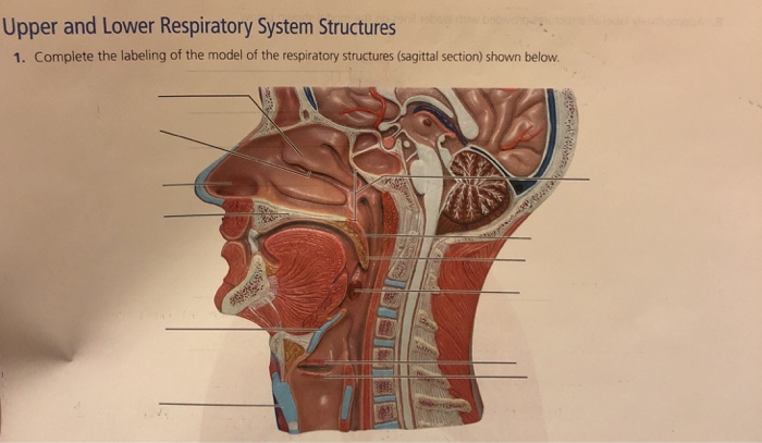
39 complete the labeling of the diagram of the upper respiratory structures
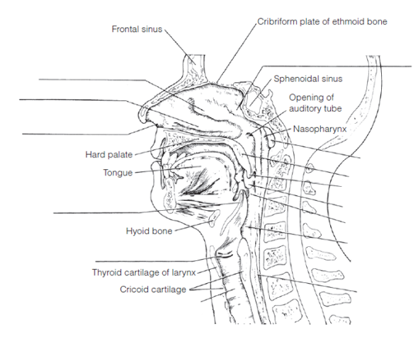
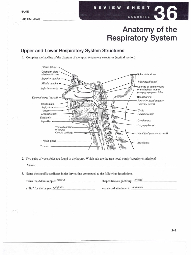
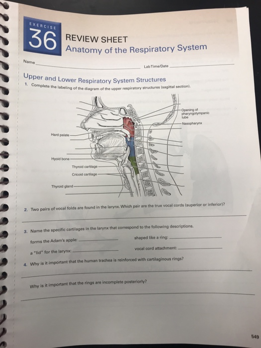
Respiratory System Upper and Lower Respiratory System Structures 1. Complete the labeling of the diagram of the upper respiratory structures (sagittal section). Frontal sinus DOLL cð/uc4LF Hard palate Tongue Hyoid bone Thyroid cartilage of larynx Cricoid cartilage Cribriform plate of ethmoid bone Sphenoidal sinus Opening of auditory tube Nasopharynx rù0S/1— escP¿fpcœs 2. What is the ... Complete the labeling of the diagram of the upper respiratory structures sagittal section. Drag the labels onto the diagram to identify the major components of the respiratory system. Part a drag the labels onto the diagram to identify features of cell signaling and receptors. In this interactive you can label parts of the human heart. Drag the ...
Complete the labeling of the diagram of the upper respiratory structures (sagittal section) Two pairs of vocal folds are found in the larynx. Which pair are the true vocal cords (superior or inferior)?
Complete the labeling of the diagram of the upper respiratory structures
Complete the labeling of the diagram of the upper respiratory structures sagittal section. Transurethral Instillation Procedure In Adult Male Mouse Protocol The kidney and urinary systems help the body to get rid of liquid waste called urea. Complete the labeling of the diagram to correctly identify the urinary system organs. Gross internal ... Complete the labeling of the diagram of the upper respiratory structures. Step 1 of 5 the respiratory tract is divided into two parts. Nose and mouth air enters nasal cavity larynx both air and food move through trachea bronchi large tubes leading to both lungs lungs. A p ii lab practical 2 review. Anatomy of the exercise36 respiratory system. Anatomy of blood vessels conduction system of the ... Upper and Lower Respiratory System Structures 1. Complete the labeling of the diagram of the upper respiratory structures (sagittal section). Frontal sinus~ Cribriform plate of ethmoid bone. Iliil'l~\ Sphenoidal. sinus. Superior Middle. concha concha ~oo. W~\\~" ~
Complete the labeling of the diagram of the upper respiratory structures. Complete the labeling of the diagram of the upper respiratory structures. Blood pressure pulse determinations anatomy of the respiratory system respiratory system physiology. Nose and mouth air enters nasal cavity larynx both air and food move through trachea bronchi large tubes leading to both lungs lungs. They are upper respiratory tract and lower respiratory tract. Step 1 of 5 the ... Upper Respiratory Tract Structural and Functional Anatomy Nose and Nasal Cavity. The nostrils, the two round or oval holes below the external nose, are the primary entrance into the human respiratory system [5].Lying just after the nostrils are the two nasal cavities, lined with mucous membrane, and tiny hair-like projections called cilia [6].During inhalation, the air passes into the nasal ... Lungs are a pair of respiratory organs situated in a thoracic cavity. Right and left lung are separated by the mediastinum. Texture-- Spongy Color - Young - brown Adults -- mottled black due to deposition of carbon particles Weight-Right lung - 600 gms Left lung - 550 gms Respiratory System Labeling - Biology Game. Identify and label figures in Turtle Diary's fun online game, Respiratory System Labeling! Drag given words to the correct blanks to complete the labeling!
Upper and Lower Respiratory System Structures. 1. Complete the labeling of the diagram of the upper respiratory structures (sagittal section).4 pages The upper respiratory system, or upper respiratory tract, consists of the nose and nasal cavity, the pharynx, and the larynx. These structures allow us to breathe and speak. They warm and clean the air we inhale: mucous membranes lining upper respiratory structures trap some foreign particles, including smoke and other pollutants, before the ... Respiratory System SHEET Upper and Lower Respiratory System Structures 1. Complete the labeling of the model of the respiratory structures (sagittal section) shown below. Nasal "Conchal Nasal meatus REVIEW as a distibule Hard plate -posterior nasal aperature -s of t palate "uvula -palentine tonsil epiglottis vestibular fold vocal fold Thyroid ... Take a look at the labeled diagram of the ... Complete the labeling of the diagram of the upper respiratory structures sagittal section. Complete the labeling of the diagram of the upper respiratory structures. Drag the labels onto the diagram to identify the structures of the upper respiratory system. 53 if 3an c t. 100 5 ratings.
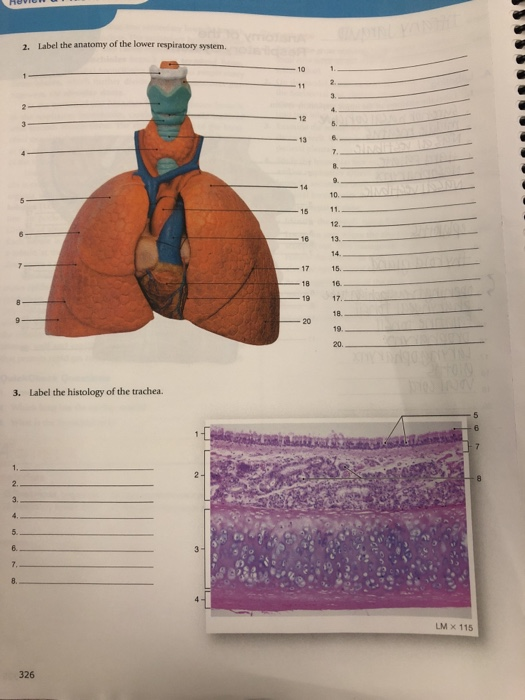
Be able to label the diagram of the upper respiratory structures (Ex 36 ... to label the diagram (figure 36.3) of the lower respiratory tract structures (Ex ... 10 Jan 2014 — Labeled diagram of the lungs/respiratory system.: --small a 1125 by 1408 pixel PNG. View Notes - Resp review from ANATOMY 1409 at Marquette University. NAME _ LAB TIME/DATE Anatomy of the Respiratory System Upper and Lower Respiratory System Structures 1. Complete the labeling of Complete the labeling of the diagram of the upper respiratory structures sagittal section two pairs of vocal folds are found in the larynx. Anatomy Of The Respiratory System It is formed by 9 supportive cartilages intrinsic and extrinsic muscles and a mucous membrane lining. Complete the labeling of the diagram of the upper respiratory structures sagittal section . Composed of the nose the ...
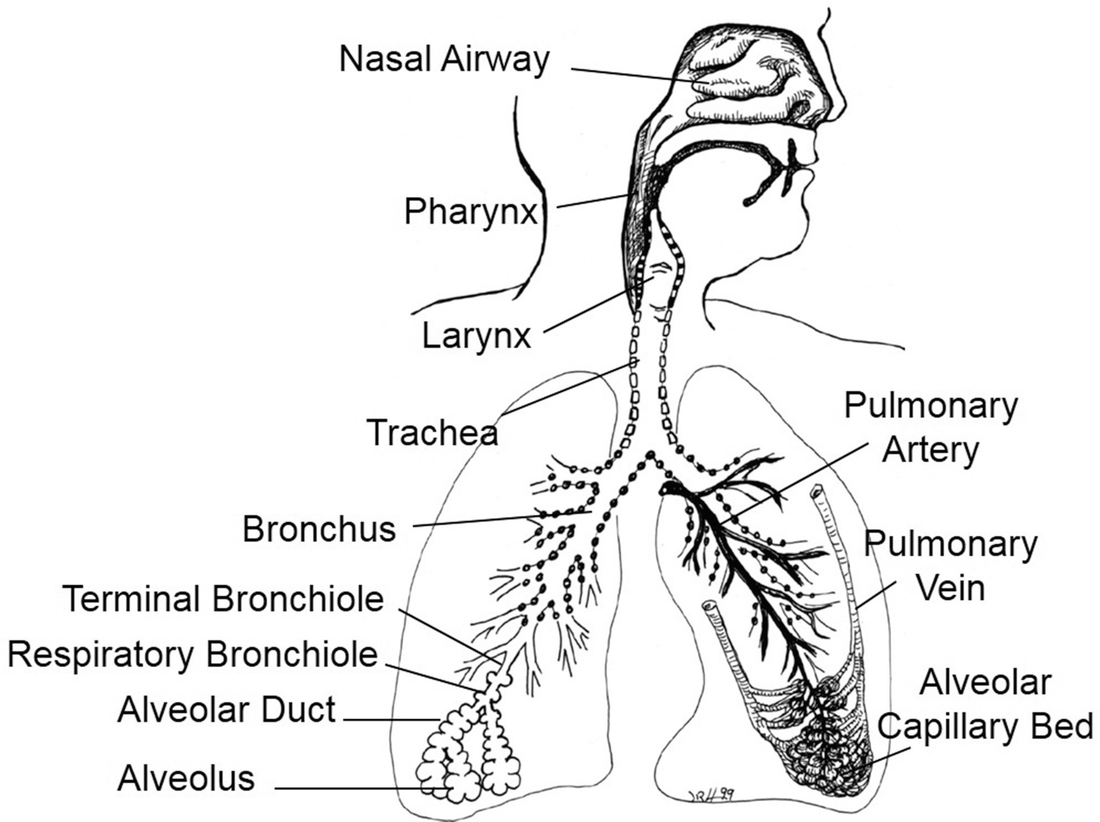
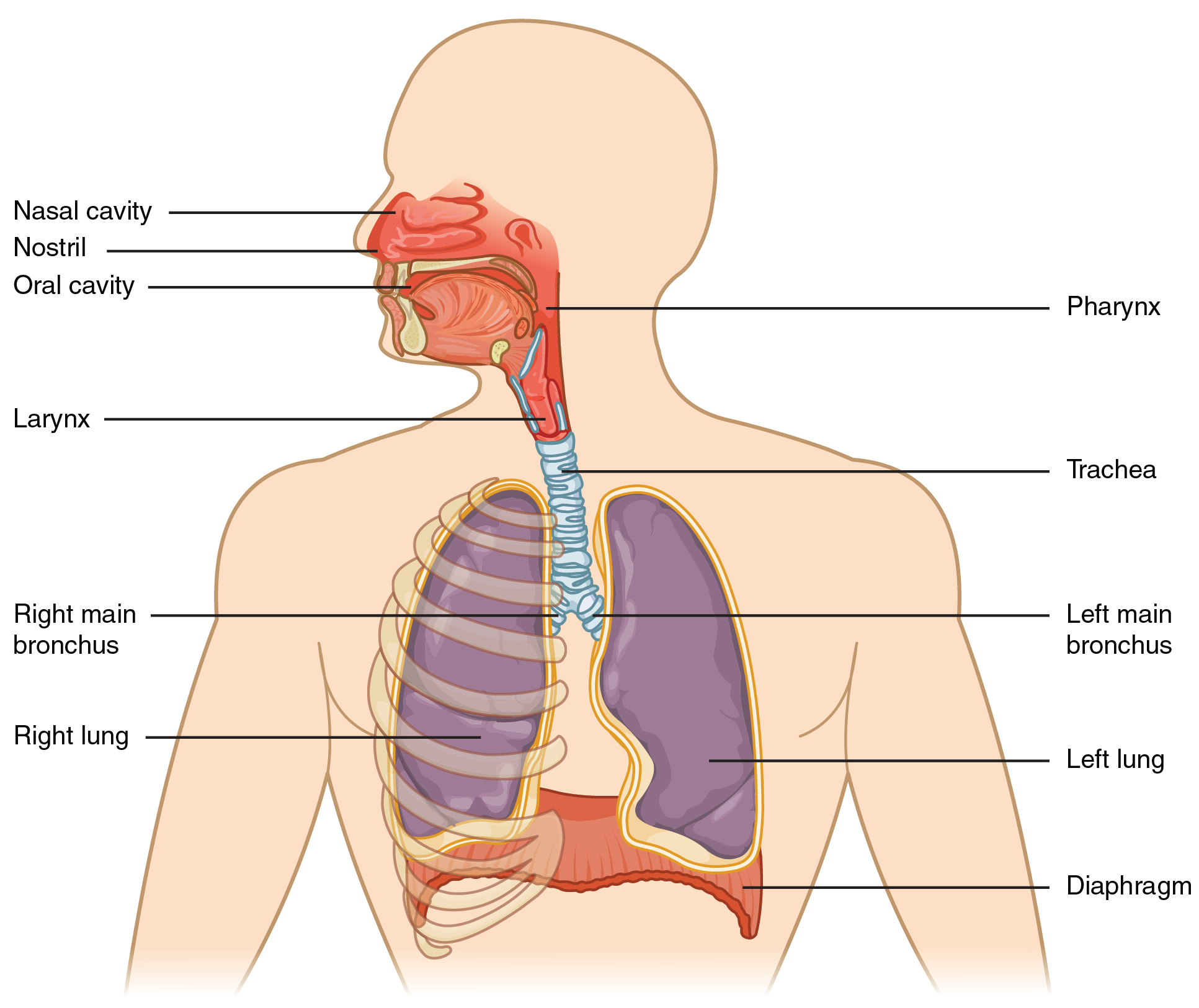
Figure 22.1.1 - Major Respiratory Structures: The major respiratory structures span the nasal cavity to the diaphragm. Functionally, the respiratory system can be divided into a conducting zone and a respiratory zone. The conducting zone of the respiratory system includes the organs and structures not directly involved in gas exchange.
The Upper Respiratory System •The following is the pathway of air: ... Figure 24.4a Respiratory Structures in the Head and Neck, Part II A sagittal section of the head and neck ... complete ring •Connecting one cartilage ring to another are annular ligaments
Take a look at the labeled diagram of the respiratory system above. As you can see, there are several structures to learn. Spend a few minutes reviewing the name and location of each one, then try testing your knowledge by filling in your own diagram of the respiratory system (unlabeled) using the PDF download below.
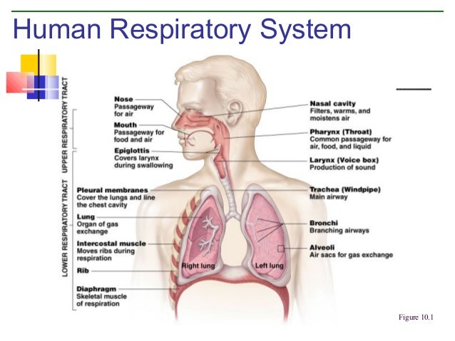
The respiratory tract is divided into two parts. They are upper respiratory tract and lower respiratory tract. Upper respiratory tract includes various passages and structures such as nose, nasal cavity, mouth, throat, and larynx (voice box). The entire respiratory tract is lined with a mucous membrane that secretes mucus.
It is formed by 9 supportive cartilages, intrinsic and extrinsic muscles and a mucous membrane lining. It is a short inch tube that is located in the throat, inferior to the hyoid bone and tongue and anterior to the esophagus. Complete the labeling of the diagram of the upper respiratory structures (sagittal section). 2.
• List the basic functions of the respiratory system. • Differentiate between respiration and ventilation. • Name and explain the functions of the structures of the upper and lower respiratory tracts. • Discuss the process of gas exchange at the alveolar level. • Describe the role of the pleura in protecting the lungs.
Complete the labeling of the diagram of the upper respiratory structures (sagittal section). 2. Two pairs of vocal folds are found in the larynx. Which pair are the true vocal cords (superior or inferior)?
Complete the labeling of the diagram of the upper respiratory structures (sagittal section).... 2. Two pairs of vocal folds are found in the larynx. Which pair are the true vocal cords (superior or inferior)? Inferior. 3. Name the specific cartilages in the larynx that correspond to the following descriptions. A.) forms the Adam's apple: B.) shaped like a ring: C.) a "lid" for the larynx: D ...
Anatomy of the respiratory system upper and lower respiratory system structures 1. Step 1 of 5 the respiratory tract is divided into two parts. Start studying ap ii review sheet 36 anatomy of the respiratory system. Complete the labeling of the diagram of the upper respiratory structures sagittal section. In the diagram at left which of the ...
View Ex26_3rd ed.pdf from BIOL 2221 at Kennesaw State University. NAM LAB TIME/DATE_ Anatomy of the Respiratory System Upper and Lower Respiratory System Structures 1. Complete the labeling of the
Structures of the Respiratory System 1. Label each of the structures indicated in this drawing of the human respiratory system. larynx trachea bronchus lung pharynx nose Gas Exchange and Transport For Questions 2-7, complete each statement by writing the correct word or words. 2. The surface area for gas exchange in the lungs is provided by ...
The respiratory tract is divided into two parts. They are upper respiratory tract and lower respiratory tract. Upper respiratory tract includes various passages and structures such as nose, nasal cavity, mouth, throat, and larynx (voice box). The entire respiratory tract is lined with a mucous membrane that secretes mucus. Chapter E23, Problem 1E is solved. View this answer View this answer ...
all respiratory passageways (conducting zone structures), besides respiratory zone structures, from nasal cavity to terminal bronchioles. why = they have no exchange function. 29. define external respiration. the gas exchange between the blood and the air-filled chambers of the lungs (oxygen loading/carbon dioxide unloading) 30.
Intact structures are shown on the left; respiratory passages are shown on the right. Select a different color for each of the structures listed below and use it to color in the coding circles and the corresponding structures on the figure. Then complete the figure by labeling the areas/structures that are provided with leader lines on the figure.
They are upper respiratory tract and lower respiratory tract. Upper respiratory tract includes various passages and structures such as nose, nasal cavity, mouth ...
Upper respiratory tract includes various passages and structures such as nose nasal cavity mouth throat and larynx voice box. Anatomy of the respiratory system upper and lower respiratory system structures 1. Complete the labeling of the diagram of the upper respiratory structures sagittal section. Two pairs of vocal folds are found in the larynx.
Upper and Lower Respiratory System Structures 1. Complete the labeling of the diagram of the upper respiratory structures (sagittal section). Frontal sinus~ Cribriform plate of ethmoid bone. Iliil'l~\ Sphenoidal. sinus. Superior Middle. concha concha ~oo. W~\\~" ~
Complete the labeling of the diagram of the upper respiratory structures. Step 1 of 5 the respiratory tract is divided into two parts. Nose and mouth air enters nasal cavity larynx both air and food move through trachea bronchi large tubes leading to both lungs lungs. A p ii lab practical 2 review. Anatomy of the exercise36 respiratory system. Anatomy of blood vessels conduction system of the ...
Complete the labeling of the diagram of the upper respiratory structures sagittal section. Transurethral Instillation Procedure In Adult Male Mouse Protocol The kidney and urinary systems help the body to get rid of liquid waste called urea. Complete the labeling of the diagram to correctly identify the urinary system organs. Gross internal ...












0 Response to "39 complete the labeling of the diagram of the upper respiratory structures"
Post a Comment